Synonyms: Ostiomeatal unit, Osteomeatal complex, OMC
Definition: The term “ostiomeatal unit” represents the area on the lateral nasal wall (middle meatus) that receives drainage from the anterior and medial ethmoid cells, frontal sinus, and maxillary sinus. It is an antomically constricted area that is prone to blockage, especially in the presence of structural anomalies, mucosal swelling or tumors. In addition, ostia themselves are small. An impairment in the ventilation of sinus due to such reasons lead to Chronic rhinosinusitis (CRS).
Boundaries:
- Medially: Middle turbinate
- Laterally: Lamina papyracea
- Superiorly and posteriorly: Basal lamella
- Inferiorly and anteriorly: It is open
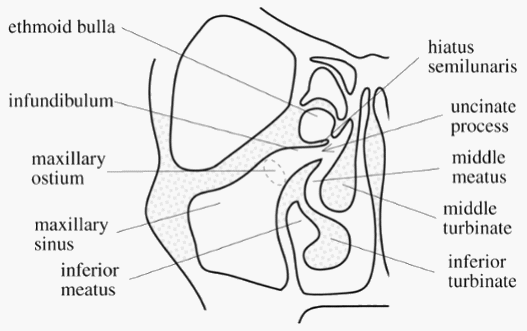
Components: Functionally, significant structures of the Ostiomeatal complex are:
- Uncinate process (Hook like bony extension of medial wall)
- Hiatus semilunaris (Crescent passage between uncinate process and ethmoid bulla through which middle meatus communicates with ethmoidal infundibulum)
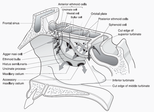
- Frontal recess (Drainage channel of frontal sinus)
- Bulla ethmoidalis (most constant and largest anterior ethmoid air cell that projects inferomedially over hiatus semilunaris)
- Ethmoidal infundibulum (Funnel shaped passage through which anterior ethmoid cells and maxillary sinus drains into middle meatus)
- Maxillary sinus ostium (Drainage channel of maxillary sinus)
Anatomic variations:
- Deviated Nasal Septum
- Concha bullosa (Enlarged, pneumatized middle turbinate
- Intralamellar cell (Air cell witihin vertical portion of middle turbinate)
- Paradoxical middle concha (Convexity of turbinate directed towards lateral nasal wall)
- Haller cells (Infraorbital ethmoid air cells)
- Agger Nasi cells (Extension of anterior ethmoid air cells into lacrimal bone)
- Uncinate process bulla
- Deviation of uncinate process
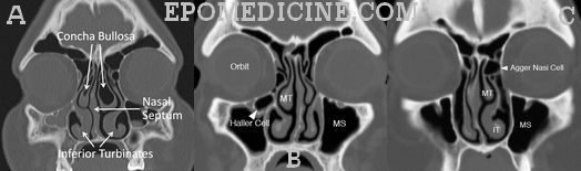
B: Haller cells
C: Agger Nasi cells
Silent sinus syndrome: Maxiallry sinus atelectasis due to chronic occlusion of maxillary sinus ostia, resulting in inward bowing of all 4 walls of the sinus including the orbital floor and increased orbital volume, leading to enophthalmos and hypoglobus (downward displacement of eye in orbit).
Note: According to some authors, basal lamella separates the anterior ostiomeatal complex (the one discussed above) and the posterior osteomeatal complex which is located in the spheno-ethmoidal recess and drains the posterior ethmoid and sphenoid sinuses. The posterior OMC is less involved in chronic sinusitis than the anterior one because its anatomic variations are fewer. So, when the disease is limited to the anterior OMC, basal lamella can be left undisturbed which reduces postoperative complications.
Great information, thanks a lot..
i’m currently working on a study that investigates the causes of performing revision frontal sinus surgery, may the authors of this article provide me with the references they used. thank you very much in advance.
Abdulwahab Alyahya
medical intern