4-6 weeks of gestation:
- Limb buds appear and elongate
- Hip joint is a single cartilaginous analage
By 7 weeks of gestation:
- Apoptotic cleft appears between cartilaginous femur and acetabulum
- Earliest time during which a hip dislocation may occur
By 11 weeks of gestation:
- Infantile configuration of hip joint is achieved
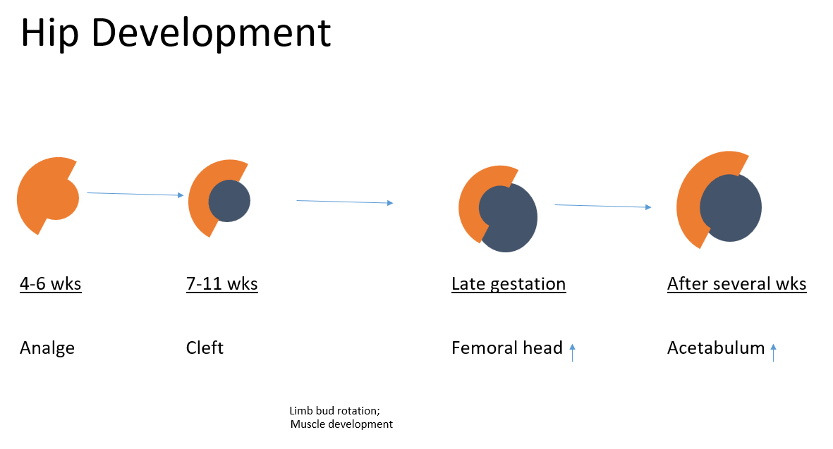
Late gestation:
- Femoral head growth is faster than acetabulum growth, resulting in under-coverage of the femoral head (<50% covered)
Postnatal:
- Acetabulum develops faster than femoral head, progressively improving the coverage
The acetabulum is deepened by the natural pressure from the developing femoral head on the acetabulum. Hips in newborns with developmental dysplasia are not just normal hips with capsular laxity; they are structurally abnormal.
Femoral anteversion: Increase with increasing fetal age, measuring on average 45 degrees at 36 weeks. Femoral anteversion then decreases in postnatal development.
Neck-shaft angle: Neck–shaft angle in fetal development appears to decrease with fetal age, ranging from approximately 145 at 15 weeks to 130 at 36 weeks. Following birth, the neck–shaft angle progressively decreases with age.
References:
- Lee MC, Eberson CP. Growth and development of the child’s hip. Orthop Clin North Am. 2006 Apr;37(2):119-32, v. doi: 10.1016/j.ocl.2005.12.001. PMID: 16638443.
- Modern Hip Preservation – New Insights In Pathophysiology And Surgical Treatment (Reinhold Ganz)
- Embryology of the Hip : Wheeless’ Textbook of Orthopaedics

He is the section editor of Orthopedics in Epomedicine. He searches for and share simpler ways to make complicated medical topics simple. He also loves writing poetry, listening and playing music. He is currently pursuing Fellowship in Hip, Pelvi-acetabulum and Arthroplasty at B&B Hospital.