Definition: Transiliac fracture dislocation of the SI joint, which combines a ligamentous disruption of the inferior part of the SI joint with a vertical fracture line of the posterior ilium, extending from the SI joint and reaching the iliac crest at different levels. The posterior superior iliac spine (PSIS) remains firmly attached to the sacrum via the superior portion of the posterior ligamentous complex.
Day Classification:
| Type | Definition | Treatment recommendations |
| I | Involves less than anterior 1/3 of SI joint – fracture line enters the SI joint at its inferior parts at the level of S2 (Outlet view); large crescent fragment | Anterolateral approach (lateral window on ilioinguinal approach): anterior SI joint plating |
| II | Involves between anterior 1/3-2/3 of SI joint – fracture line enters the SI joint between S1 & S2 foramina (Outlet view); moderate size crescent fragment | Posterior approach (Borrelli) in prone position with pelvis left free (support in chest and abdomen): lag screw + anti-glide plate |
| III | Involves more than anterior 2/3 of SI joint – fracture line enters the SI joint at the S1 level (Outlet view); small sized, superior crescent fragment | Plan A: closed/percutaneous reduction and iliosacral screw fixation – LC II channel Schanz pin can be used to control rotation – Proximal tibial traction in flexion can be used to support reduction |
| Plan B: anterior SI joint plating techniques |
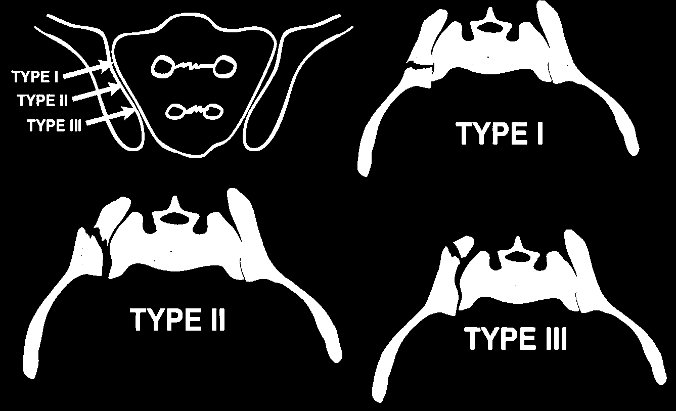
Between 12 – 30% of crescent-type fractures do not fit into the Day classification scheme.
According to the Young-Burgess classification, the crescent fractures are part of LC 2 injuries and thus considered horizontally unstable but vertically stable. In contrast, recent reports have indicated that some crescent fractures can be vertically unstable (30-40% are Tile C).
Important Surgical Considerations
1. Anterior Sacroiliac Plating:
- Protect Lateral Cutaneous Femoral Nerve (LCFN) – just below the inguinal ligament approximately 15–20 mm medial to the ASIS
- Nutrient foramen (where nutrient artery – branch from iliolumbar artery) is about 12.5 mm lateral to the SI joint
- Lumbosacral trunk is at a distance of about 10 mm lateral to the anterior SI joint
2. Posterior approach to Sacroliliac Joint:
- The localization of the nervi clunii should be considered, as the main medial branch is located approximately 8 cm lateral to the midline and 6.5 cm lateral to the PSIS at the superior iliac crest
- Exposure of the greater sciatic notch leads to optimal anatomic reduction of the SI joint
- Insertion of a curved periosteal elevator at the anterior aspect of the sacrum after dissecting the piriformis origin can protect the neurovascular bundle
- Inferior SI joint becomes visible and the anterior part of the SI joint with the anterior lateral sacral ala can be palpated
References:
- Gänsslen A, Lindahl J, Grechenig S, Füchtmeier B, editors. Pelvic ring fractures. Springer Nature; 2020 Nov 25.
- Lindahl J, Gänsslen A, Madsen JE, Krappinger D. Comparison of the AO/OTA 1996/2007 and 2018 pelvic ring fracture classifications. Arch Orthop Trauma Surg. 2024 Oct;144(10):4587-4593. doi: 10.1007/s00402-024-05557-2. Epub 2024 Oct 9. PMID: 39384630; PMCID: PMC11576778.

He is the section editor of Orthopedics in Epomedicine. He searches for and share simpler ways to make complicated medical topics simple. He also loves writing poetry, listening and playing music. He is currently pursuing Fellowship in Hip, Pelvi-acetabulum and Arthroplasty at B&B Hospital.