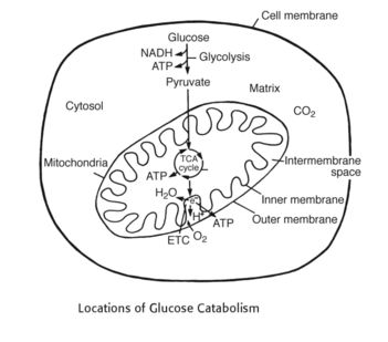
General concepts of ETC and Oxidative Phosphorylation: 1. Occurs in cell cytosol: Glycolysis 2. Occurs in mytochondrial matrix: Kreb’s (TCA) cycle 3. Occurs in mitochondrial inner membrane: ETC – Stepwise movement of electrons from high energy to low energy that activates proton pump which transports proton from the mitochondrial matrix to mitochondrial inter-membrane and…
Category: PGMEE, MRCS, USMLE, MBBS, MD/MS
Medical knowledge in bullet points with understandable language, simplified images and graspable mnemonics.
PGMEE, MRCS, USMLE, MBBS, MD/MS
Glycolysis and Gluconeogenesis: Mnemonics
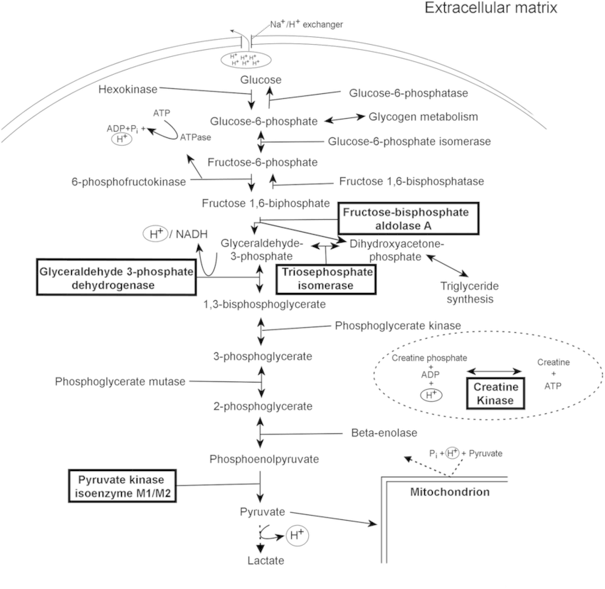
It is not necessary to memorize each and every step of the process. We will only look into the major events. A. Meaning: Glyco (Sugar) + Lysis (Breaking or splitting) B. Synonyms: Embden-Meyerhof Pathway (EM Pathway) C. Site: Cytoplasm D. Enzyme basics: Kinase: Adds or removes phosphate from substrate (uses…
PGMEE, MRCS, USMLE, MBBS, MD/MS
Kreb’s cycle (Citric Acid Cycle) : Mnemonic
Mnemonic: Our City Is Kept Safe And Sound From Malice Remember the enzymes of the cycle: All the enzymes are in the matrix of mitochondria except succinate dehydrogenase which is in inner mitochondrial membrane. Pyruvate from aerobic glycolysis enters mitochondria, where it may be converted into acetyl-CoA (irreversible reaction) under…
PGMEE, MRCS, USMLE, MBBS, MD/MS
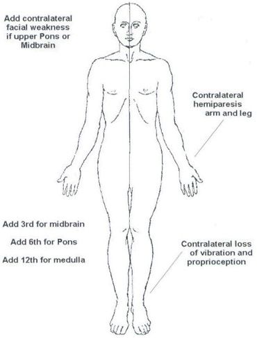
Medial Medullary (Dejerine’s) Syndrome : Anatomical basis mnemonic
As already discussed in the previous section about Lateral Medullary (Wallenberg) Syndrome: 6 “S” pass/lie on the Side (latetral) of Medulla Except the anteromedian part supplied by vertebral artery, rest of the medulla is supplied by PICA Let us now review the relevant anatomy and physiology of the medial portion…
PGMEE, MRCS, USMLE, MBBS, MD/MS
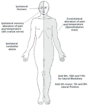
Anatomical basis of Wallenberg (Lateral Medullary) Syndrome : Mnemonic
Before proceeding into the disease itself, let’s review – relevant anatomy of the medulla with a simple mnemonic. The Side (lateral) part of Medulla contains 6 “S“ 1. Spinocerebellar tract Posterior spinocerebellar tract: Ascends and enters to ipsilateral cerebellum via ipsilateral inferior cerebellar peduncle Anterior spinocerebellar tract: Ascends and enters…
PGMEE, MRCS, USMLE, MBBS, MD/MS
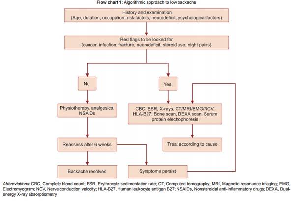
Approach to Low Back Pain
Definition of Low Back Pain Low back pain (LBP) is defined as pain, muscle tension or stiffness localized below the costal margin and above the inferior gluteal folds, with or without leg pain (sciatica). “Diagnostic triage” after excluding non-spinal causes of low back pain classifies LBP into 3 broad categories:…
PGMEE, MRCS, USMLE, MBBS, MD/MS
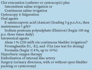
Radiation cystitis : Diagnosis, prevention and management
Cause of radiation cystitis: Bladder in the radiation field (treatment of pelvic malignancies like prostate, cervical, colorectal) Epidemiology of radiation cystitis: Incidence: 23% to 80% (variability due to variability in type and dosing of radiotherapy among different medical subspecialities) Incidence of severe hematuria: 5-8% Mean duration for developing radiation cystitis:…
PGMEE, MRCS, USMLE, MBBS, MD/MS
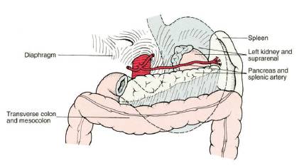
Surgical Anatomy of Stomach
GASTROESOPHAGEAL JUNCTION (CARDIA) It is the junction between esophagus and cardia of stomach Histologically: Mucosal transition from squamous to columnar epithelium Functionally: High pressure zone (Lower esophageal sphincter or LES) – Normally, LES is intraperitoneal, >2 cm long, and has a resting pressure >6 mmHg; not an anatomical sphincter but a…