Neutrophil Granules
Azurophilic (Primary) Granules
- These are lysosomes that occur in all granulocytes, as well as in lymphocytes and monocytes.
- In addition to expected lysosomal hydrolases, they also contain peroxidases (used to demonstrate azurophilic granules chemically).
- Develop earlier than specific granules.
- Stains blue/purple by Romanowsky stain.
Mnemonic: ABCDE MnOP
- Acid hydrolase
- BPI (Bactericidal Permeability Increasing) protein
- Cathepsin G
- Defensin
- Elastase
- Myeloperoxidase (MPO)
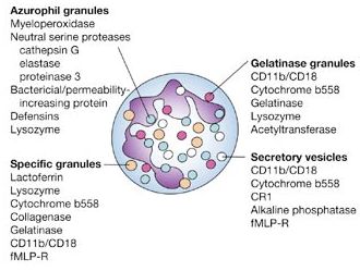
Specific (Secondary) Granules
- Do not stain intensely with Romanowsky stain.
- Smaller and more numerous than specific granules.
Mnemonic: COLA
- Collagenase
- Cathelicidin
- Oxidase (NADPH oxidase)
- Lactoferrin
- Alkaline phosphatase
Common to both Azurophilic and Specific Granules
- Lysozyme
- Phospholipase A2 (PLP A2)
Tertiary Granules
- Cathepsin
- Gelatinase
Defect in Neutrophil Granules
1. Alpha granule defect:
Myeloperoxidase (MPO) deficiency:
- Most common neutrophil defect.
- Microbicidal activity of neutrophil is delayed but not absent (defective HOCl formation).
- Acquired form is seen in AML.
Chediak-Higashi Syndrome (CHS):
- Defect in LYST (lysosomal transport) protein, encoded by CHS1 gene at 1q42.
- Abnormal giant alpha granules, impaired chemotaxis and phagolysosome formation leading to severe bacterial infections.
- Giant granules in: melanocytes and hair (oculocutaneous albinism), nerve tissue (peripheral neuropathy).
2. Specific granule defect (SGD):
- Misnomer
- Defect of both primary and specific granules: Absent specific granules + Absent Lactoferrin and ALP (primary granules) but normal MPO.
3. NADPH oxidase deficiency: Chronic Granulomatous Disease (CGD)
- Mostly X-linked recessive trait; 30% autosomal recessive
- Infections due to catalse positive organisms (organisms that destroy their own hydrogen peroxide) – extensive inflammatory reactions, and lymph node suppuration is common despite administration of appropriate antibiotics.
Platelet Granules
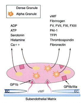
Alpha granules
Mnemonic: Alpha granules are bigger like Alpha males. These are bigger molecules like proteins and peptides. The granules can be remembered using mnemonic Platelet Function Test (PFT).
- P-selectin
- Platelet factor 4
- Platelet Derived Growth Factor (PDGF) and othr growth factors
- Fibronectin
- Fibrinogen and other factors (factor V, VIII and vWF)
- Transforming Growth Factor – beta (TGF-beta)
Delta (Dense) granules
Mnemonic: Delta granules are smaller and weakers like Delta males. These are smaller molecules. It can be remembered using mnemonic – CAN.
- Cations (Ca++, Mg++)
- Amines (Serotonin, Histamine)
- Nucleotides (ADP, ATP, PPi)
Lysosomes
Defect in Platelet Granules
Alpha granule defect:
- Gray platelet syndrome (Alpha storage pool defect) – characterized by thrombocytopenia and abnormal enlarged gray-blue platelets with a washed-out appearance due to deficeint alpha granules.
- Quebec platelet disorder – an autosomal dominant trait caused by deficient platelet alpha granule factor V (due to increased expression of alpha granule urokinase-type plasminogen activator that generates plasmin and cleaves factor V) but normal plasma factor V.
Delta granule defect:
- Delta storage pool defect – Normal number and normal appearing platelets in peripheral smear, absence of dense granules in electron microscopy and impaired second wave of platelet aggregation (prolonged bleeding time).
- Hermansky-Pudlak syndrome and Chediak-Higasghi syndrome – Rare autosomal disorders that have in common platelet dense granule deficiency, albinism and lysosomal granule defects.