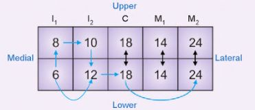
Primary teeth begin eruption at around 6 months and Permanent teeth at around 6 years. In general, lower tooth erupts before it’s upper counterpart with few exceptions (lower before upper rule). Age of 6-12 years is a period of mixed dentition (eruption of permanent 1st molar to eruption of 2nd…
Tag: Anatomy
PGMEE, MRCS, USMLE, MBBS, MD/MS
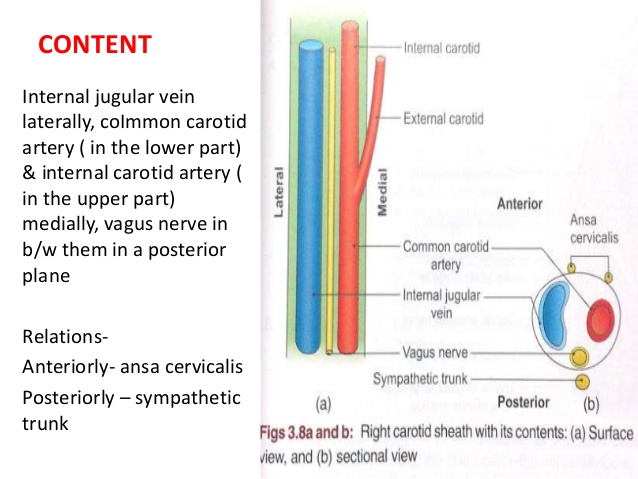
Neurovascular Relations in Anatomy
Porta hepatis Mnemonic: DAVE From anterior to posterior: Ducts (right and left hepatic duct branches) Arteries (right and left hepatic artery branches) Vein (portal vein) Epiploic foramen (foramen of Winslow) Femoral triangle or Scarpa’s triangle Mnemonic: NAVEL From lateral to medial Nerve (femoral nerve and femoral branch of genitofemoral nerve)…
PGMEE, MRCS, USMLE, MBBS, MD/MS
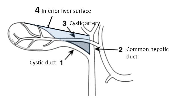
Calot’s triangle : Mnemonic
Synonyms: Calot triangle, Cystohepatic triangle Boundaries: Cysto-hepatic triangle (Budde-Rocko triangle): Calot’s triangle as described in modern surgery: Mnemonic: 3 C 1. Cystic duct 2. Common hepatic duct 3. Cystic artery Contents: Importance: The cystic artery and the duct have to be clearly defined to obtain the ‘critical view of safety’. These…
PGMEE, MRCS, USMLE, MBBS, MD/MS
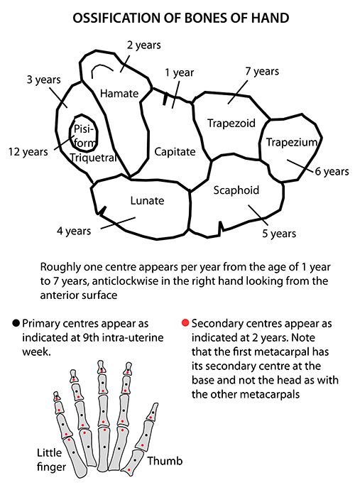
Carpal Bones Ossification: Mnemonic
Roughly one center appears per year from the age of 1 year to 7 years, anti-clockwise in right hand and clock-wise in left hand looking from the anterior surface, i.e. from ulnar side to radial side. Pisiform, being a sesamoid bone it gets left behind and only develops years later. capitate: 1-3 months hamate:…
PGMEE, MRCS, USMLE, MBBS, MD/MS
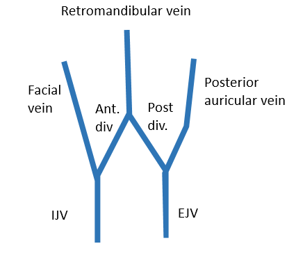
Venous drainage of Face : “W” shaped arrangement
When we look from lateral side, the veins on each side form a “W” shaped arrangement. Each corner of the “W” is prolonged upward into the scalp, and downward into the neck. Remember the 3 verticla stems of letter “W”: 1st stem (in face): Facial vein 2nd stem (behind mandible,…
PGMEE, MRCS, USMLE, MBBS, MD/MS
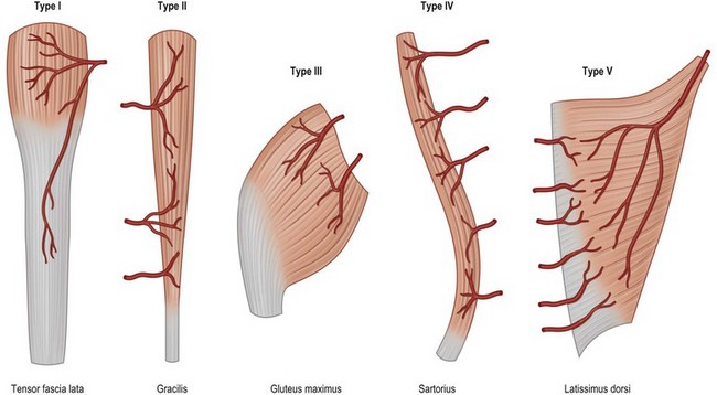
Mathes and Nahai Classification of Muscle Flap based on Vascular Anatomy
Type Dominant pedicle Minor pedicle Example I 1 – Tensor Fascia Lata (TFL) II 1 1 Gracilis III 2 – Gluteus maximus IV – Multiple Sartorius V 1 Multiple Lattisimus dorsi Mnemonic: Ten Graceful Glutes Sat on Latrines Type I: TFL Type II: Gracilis Type III: Gluteus maximus Type IV:…
PGMEE, MRCS, USMLE, MBBS, MD/MS

Axillary Artery Mnemonics
Origin of Axillary Artery: Continuation of Subclavian artery Extent of Axillary Artery: Outer border of 1st rib to Lower border of teres major (terminates as brachial artery) Relation to Axillary Vein: Lateral to Axillary Vein 3 Parts of Axillary Artery: In relation to Pectoralis Minor muscle 1st part: Proximal 2nd…
PGMEE, MRCS, USMLE, MBBS, MD/MS
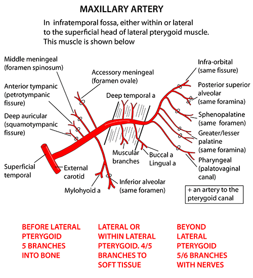
Maxillary Artery : Mnemonic
Origin of Maxillary artery: Terminal branch of External Carotid Artery (ECA) Derived from: 1st Arch Divisions of Maxillary artery: 3 parts by lateral pterygoid Branches of Maxillary artery Remember: Branches from 1st part Branches from the 2nd part Mnemonic: They supply muscles of mastication which are also derivatives of the…