Skip to content
Epomedicine

Mnemonics, Simplified Concepts & Thoughts

Epomedicine

Mnemonics, Simplified Concepts & Thoughts

venous draianage of face schematic

Venous drainage of Face : “W” shaped arrangement

Epomedicine, Oct 18, 2017

When we look from lateral side, the veins on each side form a “W” shaped arrangement.

Each corner of the “W” is prolonged upward into the scalp, and downward into the neck. Remember the 3 verticla stems of letter “W”:

  • 1st stem (in face): Facial vein
  • 2nd stem (behind mandible, in front of ear): Retromandibular vein
  • 3rd stem (behind ear): Posterior auricular vein

The two diagonal stems branching from the middle stem represents anterior and posterior division of retromandibular vein.

venous draianage of face schematic

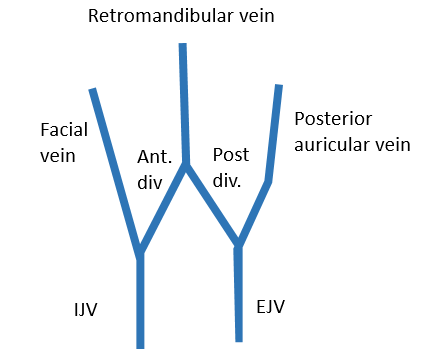
Supraorbital vein + Supratrochlear vein = Facial vein

Superficial temporal vein + Maxillary vein = Retromandibular vein

Posterior auricular vein + Posterior division of retromandibular vein = External jugular vein (EJV)

Facial vein + Anterior division of retromandibular vein = Common facial vein (drains in Internal Jugular Vein – IJV)

Deep connection of Facial vein

  1. Facial vein drains pterygoid plexus (pterygoid venous plexus drains deep veins) via deep facial vein.
  2. Pterygoid plexus has connection with cavernous sinus through the emissary veins.

How does facial vein communicate with the cavernous sinus ?

Through 3 deep connections:

  1. Superior ophthalmic vein
  2. Inferior ophthalmic vein
  3. Pterygoid plexus

Always remember!!! Facial veins are valveless.

8 shares
  • Facebook8
  • Twitter
PGMEE, MRCS, USMLE, MBBS, MD/MS AnatomyCardiovascular system

Post navigation

Previous post
Next post

Related Posts

PGMEE, MRCS, USMLE, MBBS, MD/MS

Bohr and Haldane Effect : Mnemonics

May 23, 2023May 23, 2023

Haldane effect Mnemonic: HALD Bohr Effect Mnemonic: BOHR

Read More
PGMEE, MRCS, USMLE, MBBS, MD/MS

Thoracic Diaphragm : Mnemonics

Aug 7, 2023Aug 7, 2023

Embryology Mnemonic: Several Parts Build Diaphragm Embryonic components Parts of Diaphragm 1. Septum transversum Tendinous portion (Central tendon) 2. Pleuroperitoneal folds Muscular portion 3. Body wall muscles Muscular portion 4. Dorsal mesentery of esophagus Crura 3 Major Openings of Diaphragm Mnemonics: 1. I Eat Apples 8, 10, 12 or 2….

Read More
PGMEE, MRCS, USMLE, MBBS, MD/MS internal capsule

Internal Capsule Simplified

Jul 30, 2016

Internal Capsule is a “boomerang” shaped (on horizontal section) and “funnel” shaped, i.e. tapering from superior to inferior (on sagittal section) white matter structure sandwiched between: Medially: Head of Caudate nucleus and Thalamus Laterally: Lenticular nucleus (Globus pallidus and Putamen) Parts of Internal Capsule 1. Anterior limb: Carries fibers to…

Read More

Leave a Reply Cancel reply

Your email address will not be published. Required fields are marked *

This site uses Akismet to reduce spam. Learn how your comment data is processed.

Epomedicine. Venous drainage of Face : “W” shaped arrangement [Internet]. Epomedicine; 2017 Oct 18 [cited 2026 Jun 24]. Available from: https://epomedicine.com/medical-students/venous-drainage-face-w-shaped-arrangement/.

Pre-clinical (Basic Sciences)

Anatomy

Biochemistry

Community medicine (PSM)

Embryology

Microbiology

Pathology

Pharmacology

Physiology

Clinical Sciences

Anesthesia

Dermatology

Emergency medicine

Forensic

Internal medicine

Gynecology & Obstetrics

Oncology

Ophthalmology

Orthopedics

Otorhinolaryngology (ENT)

Pediatrics

Psychiatry

Radiology

Surgery

RSS Ask Epomedicine

  • What to study for Clinical examination in Orthopedics?
  • What is the mechanism of AVNRT?

Epomedicine weekly

  • About Epomedicine
  • Contact Us
  • Author Guidelines
  • Submit Article
  • Editorial Board
  • USMLE
  • MRCS
  • Thesis
©2026 Epomedicine | WordPress Theme by SuperbThemes