Skip to content
Epomedicine

Mnemonics, Simplified Concepts & Thoughts

Epomedicine

Mnemonics, Simplified Concepts & Thoughts

Intramedullary Nail Dynamization

Dr. Sulabh Kumar Shrestha, MS Orthopedics, Apr 29, 2025Apr 29, 2025

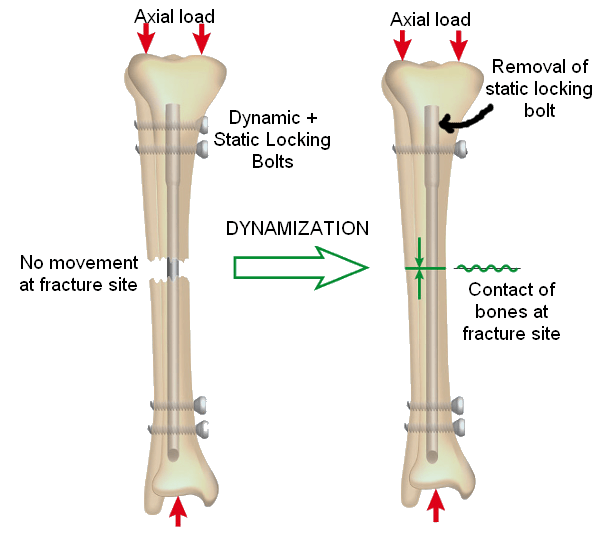
Dynamization of intramedullary nail is the process of converting a static interlocking nail (provides better fixation & rotational control) to a dynamic locking nail (allows axial loading of fracture to stimulate bone healing). Dynamization converts a bridging mode of fixation to the splinting mode.

  • Static locking: nail is locked on both cortices using screw, so that there is no relative movement between bone and nail
  • Dynamic locking: nail is locked only on distal cortex using screw and the nail will just telescope through the proximal medulla till the fracture fragments are brought together

Timing of Dynamization: Dynamization should be performed when the fracture and the newly formed callus have gained enough stability to withstand axial forces without collapse, usually 2-3 months after the initial fixation. If there is any inherent risk of shortening, dynamization should be postponed.

Indication of Dynamization: It is no longer a standard practice to dynamize the statically locked intramedullary nail if the healing is progressing normally. It is indicated when there is delay in healing and there is risk of development of nonunion or in cases of established pseudoarthrosis.

Nail dynamization

Technique of Nail Dynamization:

1. Removing the proximal static locking screw (In nails with dynamic slots): In modern nails, there are 2 proximal slots – oval one for dynamic locking and round one for static locking. Removing the static locking screw proximally will allow the proximal fragment to telescope under the limits allowed by dynamic (oval) slot while providing rotational stability which is usually adequate for bone contact.

2. Removing the screws from longer segment or farthest from fracture (In nails with only static slots): Screws are removed from longer segment to maintain adequate control of the shorter fragment.

Associated risk of dynamization: Premature dynamization may cause shortening, instability and non-union.

References:

  1. The elements of fracture fixation, 4th edition – Anand J Thakur
  2. Diagnostic imaging of musculoskeletal diseases – Akbar Bonakdarpour, William R. Reinus, Jasvir S. Khurana
  3. Current Practice of Fracture Treatment – P.C. Leung
dr. sulabh kumar shrestha
Dr. Sulabh Kumar Shrestha, MS Orthopedics

He is the section editor of Orthopedics in Epomedicine. He searches for and share simpler ways to make complicated medical topics simple. He also loves writing poetry, listening and playing music. He is currently pursuing Fellowship in Hip, Pelvi-acetabulum and Arthroplasty at B&B Hospital.

  • Facebook
  • Twitter
Surgical Skills Orthopedics

Post navigation

Previous post
Next post

Related Posts

Fellowship Blog

When Locking Screws Refuse to Budge – Techniques for Removing Jammed or Stripped Screws

Dec 6, 2025Dec 6, 2025

Written on August 10, 2025 And once again, today’s case reminded me that removing orthopedic implants can be more challenging than inserting them – especially when faced with a stubborn, stripped or jammed locking screw. We operated on a 25-year-old female with a united distal radius fracture, aiming to remove…

Read More
Surgical Skills surgical blade mnemonic

Surgical blade size : Tricks to remember

Apr 17, 2020Apr 17, 2020

Surgical blade anatomy Sharp edge: Belly Tip Unsharpened ridge opposite to the edge: Spine Slot for scalpel Surgical blade numbers Here is a trick to know the surgical blades by numbers – a simple image mnemonic which will require your imagination. We will only discuss blades commonly used in our…

Read More
Fellowship Blog

Safe Surgical Dislocation of Hip

Oct 7, 2025May 18, 2026

Surgical Principles Relevant Course of MFCA Surgical Technique 1. Position: Lateral decubitus 2. Incision and approach: Traditional Kocher-Langebeck (KL) approach or Gibson interval (more anterior interval with posterior retraction of gluteus maximus muscle without violating it) 3. Trochanteric (Digastric slide) osteotomy: 4. Exposure: 5. Z-shaped Capsulotomy: 6. Hip Dislocation: 7….

Read More

Leave a Reply Cancel reply

Your email address will not be published. Required fields are marked *

This site uses Akismet to reduce spam. Learn how your comment data is processed.

Shrestha SK. Intramedullary Nail Dynamization [Internet]. Epomedicine; 2025 Apr 29 [cited 2026 Jun 13]. Available from: https://epomedicine.com/surgical-skills/intramedullary-nail-dynamization/.

Pre-clinical (Basic Sciences)

Anatomy

Biochemistry

Community medicine (PSM)

Embryology

Microbiology

Pathology

Pharmacology

Physiology

Clinical Sciences

Anesthesia

Dermatology

Emergency medicine

Forensic

Internal medicine

Gynecology & Obstetrics

Oncology

Ophthalmology

Orthopedics

Otorhinolaryngology (ENT)

Pediatrics

Psychiatry

Radiology

Surgery

RSS Ask Epomedicine

  • What to study for Clinical examination in Orthopedics?
  • What is the mechanism of AVNRT?

Epomedicine weekly

  • About Epomedicine
  • Contact Us
  • Author Guidelines
  • Submit Article
  • Editorial Board
  • USMLE
  • MRCS
  • Thesis
©2026 Epomedicine | WordPress Theme by SuperbThemes