Skip to content
Epomedicine

Mnemonics, Simplified Concepts & Thoughts

Epomedicine

Mnemonics, Simplified Concepts & Thoughts

Bilateral V-Y (Kutler) flap for finger stump closure

Epomedicine, Feb 12, 2023Feb 12, 2023

Indications

Classically, this flap is indicated in patients with transverse or volar oblique amputations.

In actuality, the patient in whom this flap is useful generally will have an amputation where there is more tissue on the radial and ulnar margins of an amputation and exposed distal phalanx.

Limitations

Generally, the flap does not allow for advancement of more than 3 to 4 mm, and there is a sagittal scar placed on the tip of the finger.
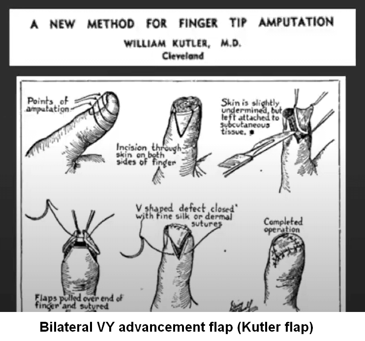
kutler flap

Technique

1. The injured area is debrided and bone spikes trimmed.

2. V-shaped incisions are made on each side of the fingertip and carried down through the subcutaneous tissue.

  • Dorsal edges of the 2 flaps begin 1-2 mm volar to the edge of the fingernail and extend volarly 7-8 mm.
  • Apex of the “V” flap sits just distal to the DIP crease.

3. Base of the triangular flap produced is held by a fine skin hook, and with sharp-pointed scissors the fibrous strands holding the skin to the bone are palpated and divided, so retaining a soft pedicle containing the neurovascular supply.

4. Flaps are sutured together over the fingertip and remaining defects are closed as a V-Y repair. However, it is not necessary to close the skin centrally over the fingertip as long as there is good soft tissue closure over the exposed distal phalanx.

Refereces:

  1. Gaber M. Kutler repair for the amputated fingertip. Ann R Coll Surg Engl. 1979 Jul;61(4):298-300. PMID: 475275; PMCID: PMC2492195.
  2. Chao, J. D., Huang, J. M., & Wiedrich, T. A. (2001). Local hand flaps. Journal of the American Society for Surgery of the Hand, 1(1), 25–44. doi:10.1053/jssh.2001.21783
  3. https://youtu.be/ZwrbhW_1mnU
  • Facebook
  • Twitter
Surgical Skills OrthopedicsPlastic surgerySurgical skills

Post navigation

Previous post
Next post

Related Posts

Surgical Skills

Krackow Suture Technique

Apr 9, 2022Dec 20, 2025

The Krackow technique is a continuous locking loop suture technique commonly used in tendon repair. The classic Krackow stitch involves 3 or more locking loops placed along each side of the ligament or tendon. Starting point: 1 cm from the tendon end Suture pitch: 5 mm (space the loops 5…

Read More
Surgical Skills

Swashbuckler (Modified Anterolateral) Approach to Distal Femur

Jul 9, 2024Jul 9, 2024

Advantages: Patient position: Supine with a roll under the knee to allow knee flexion Tourniquet: Not used unless required Incision: Superficial dissection: 1. Fascia is incised in line of incision & sharply dissected off the vastus lateralis muscle laterally to its inclusion with the iliotibial band, to expose the quadriceps…

Read More
Surgical Skills surgical blade mnemonic

Surgical blade size : Tricks to remember

Apr 17, 2020Apr 17, 2020

Surgical blade anatomy Sharp edge: Belly Tip Unsharpened ridge opposite to the edge: Spine Slot for scalpel Surgical blade numbers Here is a trick to know the surgical blades by numbers – a simple image mnemonic which will require your imagination. We will only discuss blades commonly used in our…

Read More

Leave a Reply Cancel reply

Your email address will not be published. Required fields are marked *

This site uses Akismet to reduce spam. Learn how your comment data is processed.

Epomedicine. Bilateral V-Y (Kutler) flap for finger stump closure [Internet]. Epomedicine; 2023 Feb 12 [cited 2026 Jul 10]. Available from: https://epomedicine.com/surgical-skills/bilateral-v-y-kutler-flap-for-finger-stump-closure/.

Pre-clinical (Basic Sciences)

Anatomy

Biochemistry

Community medicine (PSM)

Embryology

Microbiology

Pathology

Pharmacology

Physiology

Clinical Sciences

Anesthesia

Dermatology

Emergency medicine

Forensic

Internal medicine

Gynecology & Obstetrics

Oncology

Ophthalmology

Orthopedics

Otorhinolaryngology (ENT)

Pediatrics

Psychiatry

Radiology

Surgery

RSS Ask Epomedicine

  • What to study for Clinical examination in Orthopedics?
  • What is the mechanism of AVNRT?

Epomedicine weekly

  • About Epomedicine
  • Contact Us
  • Author Guidelines
  • Submit Article
  • Editorial Board
  • USMLE
  • MRCS
  • Thesis
©2026 Epomedicine | WordPress Theme by SuperbThemes