Skip to content
Epomedicine

Mnemonics, Simplified Concepts & Thoughts

Epomedicine

Mnemonics, Simplified Concepts & Thoughts

Wound Healing : Stages

Epomedicine, Aug 5, 2020Aug 5, 2020
wound healing phases

1. Haemostasis (immediate): In response to exposed collagen, platelets aggregate at the wound and degranulate, releasing inflammatory mediators. Clotting and complement cascades activated. Thrombus  formation and reactive vasospasm achieve haemostasis.

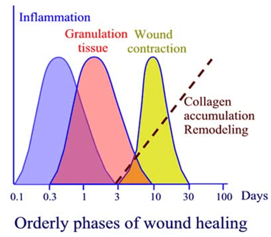
2. Inflammation (0-3 days): Vasodilatation and increased capillary permeability allow inflammatory cells to enter wound, and cause swelling. Neutrophils amplify inflammatory response by release of cytokines; reduce infection by bacterial killing; and debride damaged tissue. Macrophages follow and secrete cytokines, growth factors, and collagenases. They phagocytose bacteria and dead tissue and orchestrate fibroblast migration, proliferation, and collagen production.

3. Proliferation (3 days-3 weeks): Fibroblasts migrate into the wound and synthesize collagen. Specialized myofibroblasts containing actin cause wound contraction. Angiogenesis is stimulated by hypoxia and cytokines and granulation tissue forms

4. Remodelling (3 weeks-1 year): Re-orientation and maturation of collagen fibres increases wound strength.

Reference: Oxford Handbook of Clinical Surgery

Points to remember:
Maximum collagen production occurs at 20 days
Maximum wound strength at 3 to 6 months

  • Facebook
  • Twitter
PGMEE, MRCS, USMLE, MBBS, MD/MS General SurgeryPathology

Post navigation

Previous post
Next post

Related Posts

PGMEE, MRCS, USMLE, MBBS, MD/MS

Severity Scores for Acute Pancreatitis : Mnemonics

May 20, 2024May 20, 2024

Modified Glasgow-Imrie Score Severe pancreatitis likely: 3 or more of the following within 48 hours of symptom onset Mnemonic: PANCREAS Mnemonic Criteria Positive when P PaO2 <60 mmHg A Age >55 years N Neutrophils (WBC) >15,000/cu.mm C Calcium <2 mmol/l R RFT (Urea) >16 mmol/l E Enzyme (LDH/AST) LDH >600…

Read More
PGMEE, MRCS, USMLE, MBBS, MD/MS retroperitoneal-structures

Retroperitoneal Organs : Mnemonic

Jun 27, 2018May 13, 2024

Retroperitoneal organs are partly covered on one side with parietal peritoneum. They are immobile or fixed. The classification of retroperitoneal organs divides primary and secondary retroperitoneal organs due to the embryonic development – Retroperitoneal structures Mnemonic: SAD PUCKER Primary retroperitoneal structures Mnemonic: SAUKER Secondary retroperitoneal structures Clinical significance Kocher maneuver…

Read More
PGMEE, MRCS, USMLE, MBBS, MD/MS Heart Sounds - Murmurs Cover Picture

Heart Sounds – Murmurs

Feb 12, 2020Aug 17, 2020

Murmur is a low, indistinct sound. In terms of medicine, it is the sound produced due to turbulent flow within the heart and great vessels. They are described and named in relation to the normal heart sounds, location, and quality. Please go through these articles before reading further: Heart Sounds…

Read More

Leave a Reply Cancel reply

Your email address will not be published. Required fields are marked *

This site uses Akismet to reduce spam. Learn how your comment data is processed.

Pre-clinical (Basic Sciences)

Anatomy

Biochemistry

Community medicine (PSM)

Embryology

Microbiology

Pathology

Pharmacology

Physiology

Clinical Sciences

Anesthesia

Dermatology

Emergency medicine

Forensic

Internal medicine

Gynecology & Obstetrics

Oncology

Ophthalmology

Orthopedics

Otorhinolaryngology (ENT)

Pediatrics

Psychiatry

Radiology

Surgery

RSS Ask Epomedicine

  • What to study for Clinical examination in Orthopedics?
  • What is the mechanism of AVNRT?

Epomedicine weekly

  • About Epomedicine
  • Contact Us
  • Author Guidelines
  • Submit Article
  • Editorial Board
  • USMLE
  • MRCS
  • Thesis
©2026 Epomedicine | WordPress Theme by SuperbThemes