Slipped Capital Femoral Epiphysis (SCFE)
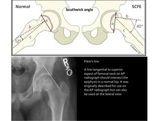
Below: Klein’s line
Southwick’s angle: Epiphyseal-shaft angle on lateral radiograph
- <30 degrees: mild
- 30-60 degrees: moderate
- >60 degrees: severe
Klein’s line: Line drawn along superior border of femoral neck (AP x-ray)
- In SCFE: will intersect less of the femoral head or not at all
Developmental Dysplasia of Hip (DDH)
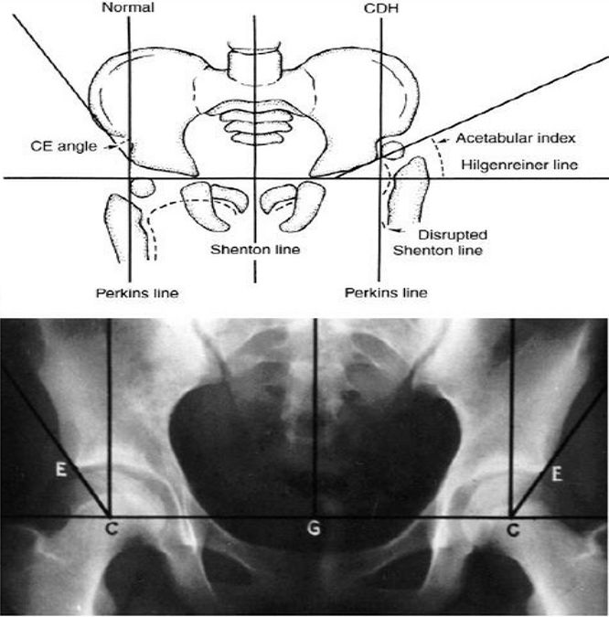
Hilgenreiner’s line: Horizontal line through triradiate cartilage of acetabulum – femoral head ossification must be below this line
Perkin’s line: Vertical line (perpendicular to Hilgenreiner’s line) from the lateral margin of ossified acetabular roof – femoral head ossification must be medial to this line
Shenton’s line: Smooth curved line connecting medial border of femoral metaphysis with superior border of obturator foramen – arc line should be continuous
Acetabular index: Angle that acetabular line (drawn from acetabular surface) makes with Hilgenreiner’s line – should be <25 degrees in >6 months old
Center edge angle of Wiberg: Angle between vertical line from center of femoral head to and line between edge of acetabulum to center of femoral head – <20 degrees is considered abnormal (reliable only in >5 year old)
In DDH:
1. Femoral head is above Hilgenreiner’s line and lateral to Perkin’s line
2. Shenton’s line is broken
3. Acetabular index increased
4. Center edge angle of Wiberg decreased
Coxa Vara
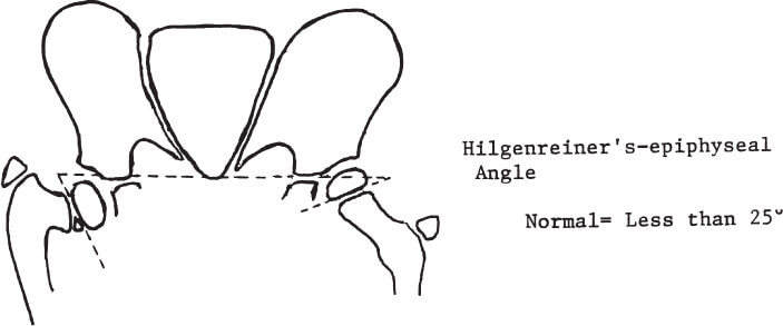
Hilgenreiner-epiphyseal angel: Angle between Hilgenreiner’s line and a line drawn parallel to capital femoral epiphysis (Increased in Coxa Vara)
Neck-shaft angle: Normal is 127 degrees (<120 degrees is Coxa Vara)
Congenital Talipes Equinovarus (CTEV)
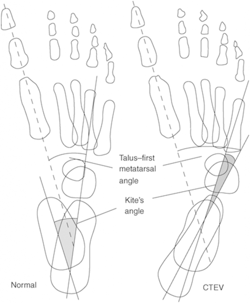
Kite’s angle (AP Talo-calcaneal angle): Angle between lines drawn down the axis of Talus and Calcaenus
- Normal: 20-40 degrees
- CTEV: <20 degrees
Talus-first metatarsal angle: Normal is 0-20 degrees (Negative in CTEV)
Calcaneal fracture
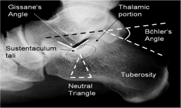
Bohler’s angle: Angle between line drawn from the highest point of anterior process of calcaneus to the highest point of posterior facet AND a line drawn tangentially from posterior facet highest point to tuberosity superior edge (normal is 20-40 degrees)
- Decreased in calcaneal fracture
Gissane’s angle: Angle formed by the downward and upward slopes of the calcaneal superior surface (normal is 120-145 degrees)
- Increased in calcaneal fracture
Ward’s Neutral triangle: The vast majority of calcaneus fracture lines propagate through a hypodense region of bone called Ward’s neutral triangle.
Supracondylar fracture of humerus
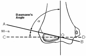
Bauman’s angle (Humero-capitellar angle): Angle between line parallel to longitudinal axis of humerus and line along lateral epiphysis (Normal 70-75 degrees; best to compare with contralateral side)
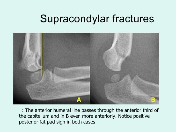
Anterior humeral line: Line drawn along anterior surface of humerus cortex must pass through the middle third of capitellum
Scoliosis
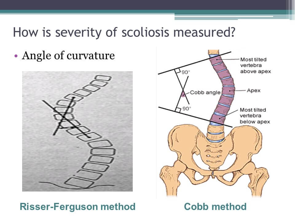
Cobb’s angle: A scoliosis is defined as a lateral spinal curvature with a Cobb angle of 10° or more
Other angles, lines and indices
Singh’s index: Grading of osteoporosis by quantifying trabeculae in neck of femur
Ward’s triangle: Femoral neck
Babcock’s triangle: Femoral neck
Fairbank’s triangle: Congenital coxa vara, Perthes disease, Nonunion neck of femur
Pauwel’s angle: Neck of femur fracture
Meary’s angle and calcaneal pitch: Pes planus and cavus
Distal radial indices: Radial length, volar tilt, ulnar variance
Gilula lines: Congruent arcs in normal wrist X-rays
Matta’s roof arc angle: Acetabular fractures
Alpha angle: Femoro-acetabular impingement
Metaphyseal-Diaphyseal angle of Drennan: Blount’s disease