Auscultation (to listen), a clinical procedure done on a daily basis as an aid in diagnosing many disorders, dates back centuries since the Egyptians. It was then an unaided auscultation (i.e. directly applying the ear to the body). In 1800’s Dr. Rene Laennec introduced the “Stethoscope” (Chest Scope), rolled paper which funneled the sounds from the chest wall to the ear. Later in 1900’s, was devised the modern stethoscope, by Dr. David Littmann.
Sounds produced in the body caught by the stethoscope:
- Heart Sounds.
- Lung Sounds.
- Bowel Sounds.
- Arterial (for measurement of Blood Pressure) and Venous Sounds.
Any sound in the circulation is created due to:
- Constriction of the blood vessel (due to turbulent flow as the principle used in measuring Blood Pressure, Arterial Bruit or Venous Hum)
- Closure of valves (as heard over the precordium from a normal Heart)
- Flow of blood through a defect in the valve or wall (as in Valvular or Septal Diseases hears as Murmurs)
- Rubbing of two surfaces (as in Pericardial Diseases hears as Pericardial Rub).
The standard description of Heart Sounds, “LUB” “DUB” are not merely two sounds, but divide the function of heart into phases and when interpreted logically (with added sounds), we can get to a conclusion whether the Heart is Normal or Diseased.
Normal Heart Sounds
In a perfectly normal heart, we observe two distinct sounds and rarely two other sounds.
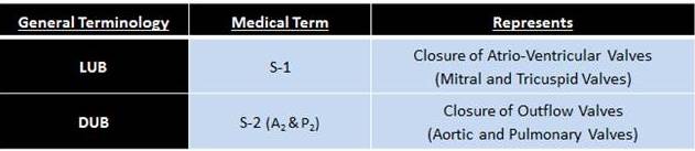
Closure of AV Valves, mark the beginning of Ventricular Systole.
Closure of Outflow Valves, mark the beginning of Ventricular Diastole.
Areas for Auscultation on the Chest
The areas of the heart sounds being heard do not represent the actual presence of the valves but are due to transmission of the sounds.
As sounds always get transmitted easily through a high pressure system, the sounds produced by the closure of valves travel through the blood which is into a High Pressure Area (which is also the direction of Blood Flow).
- Mitral Valve Closure:- Sound travels into the Left Ventricle and is heard where the ventricle wall touches the pericardium Mitral Area i.e., 5th Intercostal Space at the Mid-Clavicular Line.
- Tricuspid Valve Closure:- Sound travels into the Right ventricle and is heard where its wall touches the pericardium Tricuspid Area i.e., 4th Intercostal Space at the Left Parasternum.
- Aortic Valve Closure:- Sound travels into the Aorta (since the Left Ventricle is in Diastole) and is heard where Aorta is near to the pericardium Aortic Area i.e., 2nd Intercostal Space at the Right Parasternum.
- Pulmonary Valve Closure:- Sound travels into the Pulmonary Artery (since the Right Ventricle is in Diastole) and is heard where the Pulmonary Artery is near to the pericardium Pulmonary Area i.e., 2nd Intercostal Space at the Left Parasternum.

Areas on the Precordium for Auscultation for a specific Sound
S-1
Mitral Area (Left 5th Intercostal Space, Mid Clavicular Line).
Tricuspid Area (Left 5th Intercostal Space, Sternal Border).
M1 closes before T1 (but does not split)
S-2
Aortic Area (Right 2nd Intercostal Space, Sternal Border).
Pulmonary Area (Left 2nd Intercostal Space, Sternal Border).
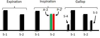
A2 heard before P2 (during Inspiration)
S-2 is heard as two sound during Inspiration known as the Spilt. This is due to:
- 1. a. During Inspiration the Pressure in the Thorax (Intra-thoracic Pressure) reduces in order to facilitate the entry of air into the lungs, as the pressure decreases, the venous return to the Right side of the Heart Increases.
- 1. b. Also during Inspiration the lung increase in volume and there occurs pooling of blood in the Pulmonary Vasculature leading to reduced filling of the Left Side of the Heart compared to the Right Side.
The increased filling of the Right Ventricle and reduced Filling of the Left Ventricle causes delay in emptying the right ventricle and hence Pulmonary Valve closes a little later than the Aortic Valve.
- 2. Left Ventricle pumps blood into High Resistance System (Systemic Circulation) in contrast to the Right Ventricle which pumps blood to a Low Resistance System (Pulmonary Circulation).
Hence right ventricle empties for a longer duration than the left causing Pulmonary Valve closing after the Aortic Valve.
S-3
Mitral Area (Left 5th Intercostal Space, Mid Clavicular Line).
Tricuspid Area (Left 5th Intercostal Space, Sternal Border).
S – 3 is heard during the Mid Diastolic Phase
S-3 is normal in people aged < 35 years.
This is due to Passive Filling of Blood when it strikes a Compliant Ventricle Wall. Adding to this, the vibrations of Atrio-ventricular Valves also contribute.
S-4
Mitral Area (Left 5th Intercostal Space, Mid Clavicular Line).
Tricuspid Area (Left 5th Intercostal Space, Sternal Border).
S – 4 is heard towards the End of Diastole
S-4 is due to Forceful Filling of Blood into a Stiff Ventricle. The Vibrations of the Atrio-Ventricular Valves also add up to a minor extent.
When S-1, S-2 , S-3 are heard, it is named as “KENTUKY RHYTHM”.
When S-1, S-2, S-4 are heard, it is named as “TENNESSEE RHYTHM”.
When S-1, S-2, S-3, S-4 are heard, it is named as “GALLOP RHYTHM”.
Summary
Heart, “The Blood Pump” produces sounds sue to the closure of valves during the Cardiac Cycle, commonly referred to as “LUB — DUB” when picked up by the Stethoscope.
In a Healthy individual, 2 sounds are heard viz. S-1 and S-2 and rarely S-3, S-4 or both may be heard.
These sounds are clinically significant in diagnosing many conditions of the Cardiac System.
References
- Harrison’s Principles of Internal Medicine, 20th Edition.
- Bates’ Guide to Physical Examination and History Taking, 10th Edition.
- Hutchinson’s Clinical Methods, 23rd Edition.
Reviewed by: Mr. L. Prakasham Reddy, Asst. Prof., Department of Physiology, Kamineni Institute of Medical Sciences.

Graduated from one of the famous institutions in Telangana, Kamineni Institute of Medical Sciences. He has always been fond of writing articles in Medicine. Since the undergraduate years was interested in making creative Presentations and taking seminars.


Great work sir. Your descriptions are very simple to understand thanks