Skip to content
Epomedicine

Mnemonics, Simplified Concepts & Thoughts

Epomedicine

Mnemonics, Simplified Concepts & Thoughts

trigeminal sensory map

Trigeminal Nerve Simplified

Epomedicine, Jul 26, 2016Oct 26, 2017

Course of Trigeminal Nerve and Trigeminothalamic Pathway

Trigeminal nerve course

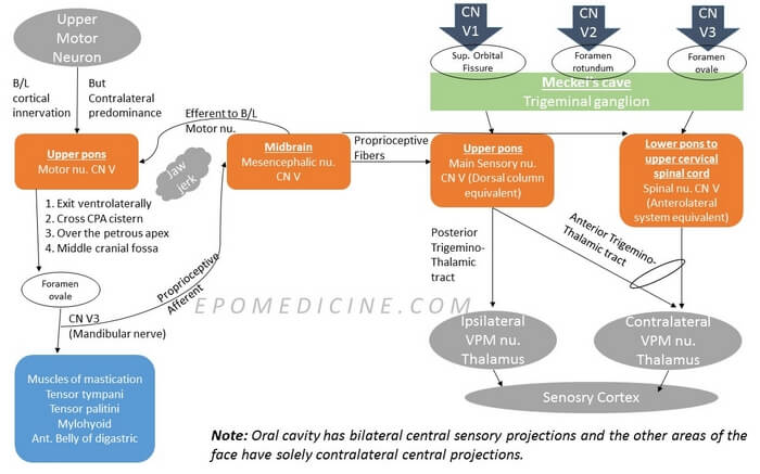
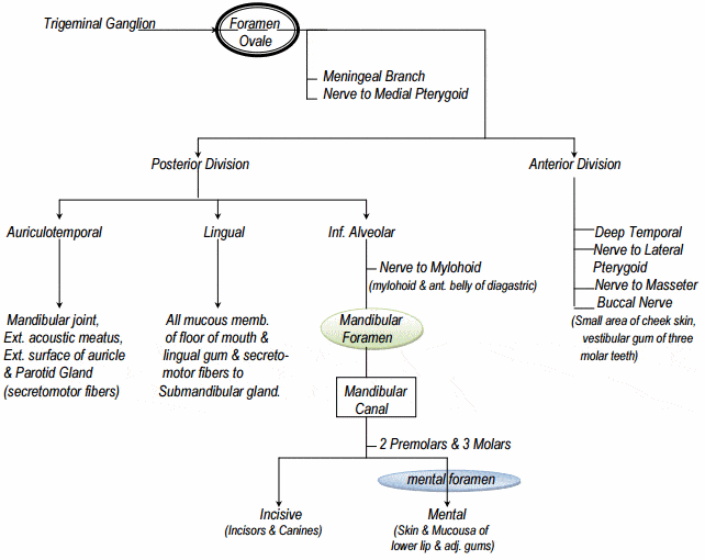
Mandibular (CN V3) Division of Trigeminal Nerve

mandibular-trigeminal

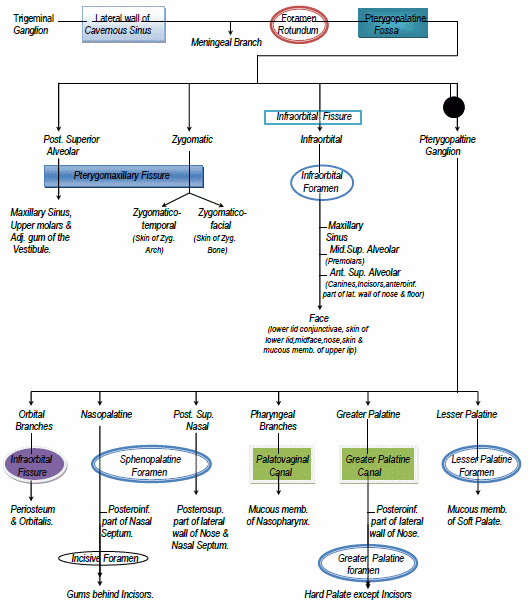
Maxillary (CN V2) Division of Trigeminal Nerve

maxillary trigeminal

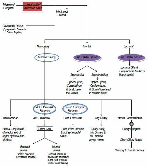
Ophthalmic (CN V1) Division of Trigeminal Nerve

ophthalmic trigeminal

Sensory Map Of Trigeminal Nerve on Face

trigeminal sensory map

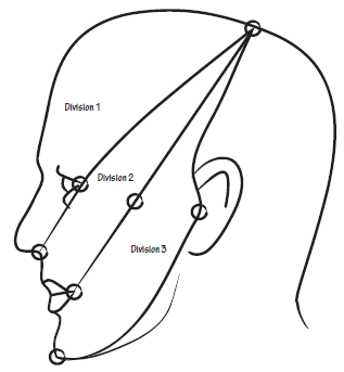
Area of Ophthalmic division: Line joining –

  1. Just behind the top of head
  2. Corner of eyes
  3. Tip of nose

Area of Maxillary division: Line joining –

  1. Same point as above in head
  2. Maxilla
  3. Angle of mouth

Area of Mandibular division: Line joining –

  1. Same point as above in head
  2. Tragus of ear
  3. Mentum

Important point to note:

Mandibular division neither covers the angle of mandible, nor the outer ear. These areas are often tested to differentiate neurologic and functional (non-neurologic symptoms) as Trigeminal nerve doesn’t have sensory supply to these regions. These regions are supplied by:

  • Outer ear: Cranial nerve VII, IX and X
  • Angle of Mandible: C2, C3

Lateral Pterygoids

While the other muscles of mastication closes the jaw, the lateral pterygoids open the jaw. Like, genioglossus of tongue, the fibers crosses and inserts to the angle of jaw on the opposite side. Hence, dysfunction of lateral pterygoid at one side leads to deviation of jaw in the same side due to the action of opposite lateral pterygoid.

In Upper Motor Neuron lesion:

  • They have bilateral cortical innervation with contralateral predominance.
  • Hence, there may be no visible deviation of jaw during clinical examination.
  • If deviation is seen: Deviation of jaw to the side contralateral to the UMN lesion
    • Deviation at one side → Dysfunction of lateral pterygoid in same side → Contralateral UMN lesion

In Lower Motor Neuron lesion:

  • Deviation of the jaw to the side ipsilateral to the LMN lesion.

Jaw Jerk

  1. Stimuli: Stretch applied by reflex hammer on relaxed jaw
  2. Sensory receptors: Muscle spindles of muscles of mastications
  3. Afferent: Proprioceptive fibers from CN V3
  4. Center: Mesencephalic nucleus of trigeminal nerve (Not the trigeminal ganglion)
  5. Efferent: Bilateral innervation to the motor nucleus of facial nerve
  6. Response: Closure of jaw
  • Facebook
  • Twitter
PGMEE, MRCS, USMLE, MBBS, MD/MS AnatomyNervous system

Post navigation

Previous post
Next post

Related Posts

PGMEE, MRCS, USMLE, MBBS, MD/MS Brain blood supply

Circle of Willis and Forebrain Blood Supply

Aug 1, 2016Aug 1, 2016

We have already discussed earlier on the intracranial course of Internal Carotid Artery (ICA) and Circle of Willis formation with the help of a simple mnemonic. General Concepts of Blood Supply of Brain and Spinal Cord 1. Spinal cord, Hind-brain and Mid-brain: Veterbro-basilar system 2. Forebrain: Circle of willis which…

Read More
PGMEE, MRCS, USMLE, MBBS, MD/MS lateral brainstem syndrome

Anatomical basis of Wallenberg (Lateral Medullary) Syndrome : Mnemonic

Jan 21, 2016Oct 26, 2022

Before proceeding into the disease itself, let’s review – relevant anatomy of the medulla with a simple mnemonic. The Side (lateral) part of Medulla contains 6 “S“ 1. Spinocerebellar tract Posterior spinocerebellar tract: Ascends and enters to ipsilateral cerebellum via ipsilateral inferior cerebellar peduncle Anterior spinocerebellar tract: Ascends and enters…

Read More
PGMEE, MRCS, USMLE, MBBS, MD/MS

Pathophysiology of Acute Compartment syndrome

Apr 27, 2023Apr 27, 2023

Definition Mubarak and Hargens (1981) defined compartment syndrome as an elevation of the interstitial pressure in a closed osseofascial compartment that results in microvascular compromise. Pathophysiology Tissue perfusion = Capillary perfusion pressure – Interstitial fluid pressure 3 major theories for microvascular dysfunction and ischemia: Tissue survival: Vicious cycle of compartment…

Read More

Leave a Reply Cancel reply

Your email address will not be published. Required fields are marked *

This site uses Akismet to reduce spam. Learn how your comment data is processed.

Pre-clinical (Basic Sciences)

Anatomy

Biochemistry

Community medicine (PSM)

Embryology

Microbiology

Pathology

Pharmacology

Physiology

Clinical Sciences

Anesthesia

Dermatology

Emergency medicine

Forensic

Internal medicine

Gynecology & Obstetrics

Oncology

Ophthalmology

Orthopedics

Otorhinolaryngology (ENT)

Pediatrics

Psychiatry

Radiology

Surgery

RSS Ask Epomedicine

  • What to study for Clinical examination in Orthopedics?
  • What is the mechanism of AVNRT?

Epomedicine weekly

  • About Epomedicine
  • Contact Us
  • Author Guidelines
  • Submit Article
  • Editorial Board
  • USMLE
  • MRCS
  • Thesis
©2026 Epomedicine | WordPress Theme by SuperbThemes