Skip to content
Epomedicine

Mnemonics, Simplified Concepts & Thoughts

Epomedicine

Mnemonics, Simplified Concepts & Thoughts

lungs lymph nodes

TNM and Staging of Lung Cancer Simplified

Epomedicine, Aug 15, 2016

The current staging system is recommended for the classification of both non–small cell and small cell lung carcinomas and for carcinoid tumors of the lung .

TNM Staging of Lung Cancer

Primary Tumor (T)

The “T” staging of bronchogenic carcinoma is a bit complex compared to others. Five things must be checked before assigning a T classification. They can be remembered with a mnemonic: ABCDE.

  1. Atelectasis
  2. Bronchus involvement (bronchoscopy)
  3. Contiguous conquer (Adjacent site invasion)
  4. Diameter in cm
  5. Elevated nodules

Chest wall involvement as in breast cancer denotes advanced T stage.

Diameter (cm) Bronchoscopy (Main bronchus involvement) Atelectasis or Obstructive pneumonia Invasion Nodules
T1 A – ≤2 cm
B – 2 to ≤3 cm
T2 A – 3 to ≤5 cm ≥2 cm distal to carina Lobar Visceral pleura
B – 5 to ≤7 cm
T3 >7 cm <2 cm distal to carina Whole lung Chest wall (including pancoast or superior sulcus tumor), Diaphragm, Phrenic nerve, Mediastinal or parietal pleura Same lung lobe
T4 Any size Carina Heart, great vessels, trachea, esophagus, recurrent laryngeal nerve spine, Different ipsilateral lung lobe

lungs lymph nodes

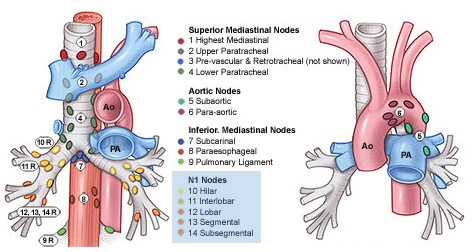
Regional Lymph Nodes (N)

  • N1: Ipsilateral – Peribronchial, hilar or intrapulmonary nodes
  • N2: Ipsilateral – Mediastinal or subcarinal nodes
  • N3:
    • Contralateral – Mediastinal or Hilar (N1 and N2 if ipsilateral)
    • Ipsilateral or Contralateral: Scalene or Supraclavicular nodes

Metastasis (M)

M1:

  • a – Separate tumor nodule in contralateral lung lobe (T4 if on ipsilateral lobe), Pleural nodules, Malignant pleural or pericardial effusion
  • b – Distant Metastasis

AJCC Staging of Lung Cancer
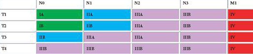
lung cancer staging matrix

The staging of lung cancer is similar to another thoracic tumor – breast cancer. In both of these sites, the primary drainage nodes (N1) is close to the involved site, hence, T1=N1.

  • M stage is a separate stage: M1 = Stage IV
  • N stage determines stage group I-III
    • N3 = Stage IIIB
    • N2 = Stage IIIA except with T4 (Stage III B)
  • T stage modifies substages:
    • T4 = Stage IIIB
    • T1 or T2 N0 = Stage IA or IB respectively
    • T1 or T2 N1 = Stage IIA or IIB respectively

Algorithm for Lung Cancer Management
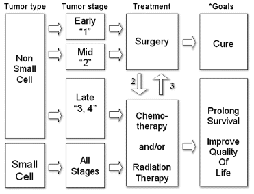
lung-cancer-treatment

39 shares
  • Facebook39
  • Twitter
PGMEE, MRCS, USMLE, MBBS, MD/MS Internal medicineOncologyRespiratory system

Post navigation

Previous post
Next post

Related Posts

PGMEE, MRCS, USMLE, MBBS, MD/MS

Metaphyseal lesions : Differential Diagnoses

Jul 25, 2024Jul 25, 2024

Lesions Age Demographics Site Presentation Imaging Treatment Comments Acute Osteomyelitis Bimodal (<2 years and 8-12 years) M:F = 2:1 Distal femur, Proximal tibia Pain, refusal to bear weight, erythema, warmth, fluctuance, tenderness Lags 2 weeks behindDestruction of bone, periosteal reaction (lamellated, hair on end), new bone formation 1. Antibiotics2. Surgical…

Read More
PGMEE, MRCS, USMLE, MBBS, MD/MS parinaud syndrome

Parinaud Syndrome – Anatomical Basis

May 19, 2019May 19, 2019

Synonyms: Dorsal midbrain sydnrome, Pretectal syndrome Main components of Parinaud syndrome: Vertical gaze palsy Convergence-retraction nystagmus Pupillary light-near dissociation Lid retraction (Collier’s sign) Anatomical basis of Parinaud syndrome: Site of lesion: Dorsal midbrain Close to cerebral aqueduct and pineal gland – hydrocephalus and pinealoma Arterial supply – posterior cerebral artery…

Read More
PGMEE, MRCS, USMLE, MBBS, MD/MS ovarian neoplasm

Pathology of Ovarian Tumors – Quick Review

Feb 7, 2017

The latest TNM and FIGO staging for Ovarian Cancer has been discussed earlier here. Here, we will discuss pathology of ovarian tumors in short and in a way thats easy to grasp. WHO Classification of Ovarian Tumors Cells of origin Surface coelomic epithelium Germ cells Sex cord, stromal cells Metastatic…

Read More

Comment

  1. OncoNorm Plus says:
    Sep 29, 2020 at 10:00 am

    Great content and information. Knowing the staging of lung cancer and its TNM is very necessary if someone is having doubts about having lung cancer.

    Reply

Leave a Reply Cancel reply

Your email address will not be published. Required fields are marked *

This site uses Akismet to reduce spam. Learn how your comment data is processed.

Epomedicine. TNM and Staging of Lung Cancer Simplified [Internet]. Epomedicine; 2016 Aug 15 [cited 2026 Sep 29]. Available from: https://epomedicine.com/medical-students/tnm-staging-lung-cancer-simplified/.

Pre-clinical (Basic Sciences)

Anatomy

Biochemistry

Community medicine (PSM)

Embryology

Microbiology

Pathology

Pharmacology

Physiology

Clinical Sciences

Anesthesia

Dermatology

Emergency medicine

Forensic

Internal medicine

Gynecology & Obstetrics

Oncology

Ophthalmology

Orthopedics

Otorhinolaryngology (ENT)

Pediatrics

Psychiatry

Radiology

Surgery

RSS feed: Ask Epomedicine Ask Epomedicine

  • What to study for Clinical examination in Orthopedics?
  • What is the mechanism of AVNRT?

Epomedicine weekly

  • About Epomedicine
  • Contact Us
  • Author Guidelines
  • Submit Article
  • Editorial Board
  • USMLE
  • MRCS
  • Thesis
©2026 Epomedicine | WordPress Theme by SuperbThemes