Synonyms: Hoffman-Tinel test, Tinel’s sign, Nerve percussion test
Definition: “pins and needle feeling” elicited by tapping on a nerve proximally, with resulting paresthesia experienced in the corresponding distal cutaneous distribution of an injured peripheral nerve.
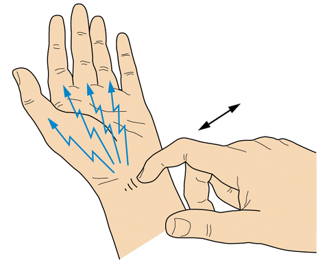
Eliciting tinel sign: With gentle percussion by a finger or percussion hammer along the course of an injured nerve. It should be tested for in a distal to-proximal direction.
Clinical significance: a provocative test that can help clinicians diagnose entrapment neuropathies as well as peripheral nerve regeneration following nerve injury. Distal progression of the response along the course of the nerve in question can be measured, and some have used the rate of this progression to establish prognosis or suggest the need for exploration.
Pathophysiology: Compression of a peripheral nerve does result in structural changes (i.e., demyelination and remyelination), which lead to various degrees of focal axonal degeneration and regeneration. These changes are believed to cause the nerve to become sensitive to mechanical stimuli such as pressure or percussion. Regenerating touch fibers are oversensitive to mechanical stimulation, and after they reach the skin they remain hypersensitive to gentle tapping or gentle stroking for some time. A positive Tinel sign is presumptive evidence that regenerating axonal sprouts that have not obtained complete myelinization are progressing along the endoneurial tube. With progressive regeneration, the positive response fades proximally, presumably because of progressive myelinization along the more proximal part of the regenerated segment.
Interpretation: A transient tingling sensation should be felt in the distribution of the injured nerve rather than at the area percussed and the sensation should persist for several seconds after stimulation. and never painful. If there is any pain associated with gentle tapping of a sensory or mixed nerve, this finding should not be called Tinel’s sign, but should be regarded as evidence of a neuroma or as a neuroma-like sign.
Tinel sign and Sunderland classification: A distally advancing Tinel sign should occur in Sunderland types 2 and 3 nerve injuries. A Sunderland type 1 injury or neurapraxia should not show an advancing Tinel sign because wallerian degeneration and axonal regeneration do not occur. A Sunderland type 4 or type 5 injury would not show an advancing Tinel sign unless repaired.
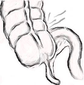
Motor tinel sign: The motor Tinel sign (MTS) refers to electromyographic activity, sometimes associated with a visible muscle jerk, evoked by percussion or manipulation of a peripheral nerve.
Tender muscle sign: Squeezing a denervated muscle a few weeks after nerve repair produces a characteristic response in patients. This response is observed before any clinical evidence of motor recovery.
References:
- Ho T, Braza ME. Hoffmann Tinel Sign. [Updated 2020 Dec 2]. In: StatPearls [Internet]. Treasure Island (FL): StatPearls Publishing; 2020 Jan-. Available from: https://www.ncbi.nlm.nih.gov/books/NBK555934/
- Sansone JM, Gatzke AM, Aslinia F, Rolak LA, Yale SH. Jules Tinel (1879-1952) and Paul Hoffman (1884-1962). Clin Med Res. 2006 Mar;4(1):85-9. doi: 10.3121/cmr.4.1.85. PMID: 16718952; PMCID: PMC1435662.
- https://citeseerx.ist.psu.edu/viewdoc/download?doi=10.1.1.899.9399&rep=rep1&type=pdf
- Campbell’s Operative Orthopedics – 13th Edition
- Montagna P, Liguori R. The motor tinel sign: a useful sign in entrapment neuropathy? Muscle Nerve. 2000 Jun;23(6):976-8. doi: 10.1002/(sici)1097-4598(200006)23:6<976::aid-mus22>3.0.co;2-6. PMID: 10842279.
- Lee EY, Karjalainen TV, Sebastin SJ, Lim AY. The value of the tender muscle sign in detecting motor recovery after peripheral nerve reconstruction. J Hand Surg Am. 2015 Mar;40(3):433-7. doi: 10.1016/j.jhsa.2014.12.018. PMID: 25708431.