Skip to content
Epomedicine

Mnemonics, Simplified Concepts & Thoughts

Epomedicine

Mnemonics, Simplified Concepts & Thoughts

Spinal Tumors Localization : Mnemonics

Epomedicine, Apr 9, 2025Apr 9, 2025

Intradural Intramedullary Spinal Tumors

Mnemonic: I HEAL

  1. Infarction
  2. Hemangioblastoma
  3. Ependymoma
  4. Astrocytoma
  5. Lipoma

Intradural Extramedullary Spinal Tumors

Mnemonic: MNM

  1. Meningioma (25%)
  2. Nerve sheath tumors (35%)
  3. Metastases
spinal tumor location
Kumar, Naresh & Tan, Wei & Wei, Wei & Vellayappan, Balamurugan. (2020). An overview of the tumors affecting the spine—inside to out. Neuro-Oncology Practice. 7. i10-i17. 10.1093/nop/npaa049.

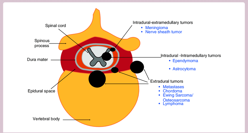
Extradural Spinal Tumors

Lesions outside the thecal sac are categorized as extradural lesions. Remember that everything that isn’t in the thecal sac is extradural, including discs, bones, nerves, and blood vessels.

1 shares
  • Facebook
  • Twitter
PGMEE, MRCS, USMLE, MBBS, MD/MS Internal medicineNervous systemOrthopedics

Post navigation

Previous post
Next post

Related Posts

PGMEE, MRCS, USMLE, MBBS, MD/MS air bronchogram

Chest X-ray: Alveolar vs Interstitial Disease

Jun 19, 2015Jun 19, 2015

Interstitium is the scaffolding that supports the alveolar walls and surrounds both the alveoli and the terminal bronchioles. Neither alveoli nor interstitium is visible on a chest X-ray when normal. It is necessary to analyze whether the pattern of diffuse opacification in the lung field is alveolar or interstitial. Terms:…

Read More
PGMEE, MRCS, USMLE, MBBS, MD/MS

How Metformin Causes Vitamin B12 deficiency?

Aug 9, 2019May 31, 2020

Multiple studies have shown that metformin reduces serum B12 levels in 10–30% of patients. The effect increases with the duration of the therapy. Decrease in vitamin B12 absorption and levels following metformin use typically starts as early as the 4th month. Mechanisms of Vitamin B12 deficiency with Metformin Use: It…

Read More
Emergency Medicine pulmonary embolism xray

Pulmonary Embolism : Chest X-ray Signs

Oct 25, 2017

1. Classic presentation is normal X-ray in patient with dyspnea and hypoxia 2. Atelectasis or parenchymal abnormality (68%) 3. Elevated hemidiaphragm 4. Pleural effusion (Felson’s sign – pleural effusion on left > right) 5. Hampton’s hump: peripheral pleural based wedge-shaped density above the diaphragm due to pulmonary infarct 6. Westermark’s…

Read More

Leave a Reply Cancel reply

Your email address will not be published. Required fields are marked *

This site uses Akismet to reduce spam. Learn how your comment data is processed.

Pre-clinical (Basic Sciences)

Anatomy

Biochemistry

Community medicine (PSM)

Embryology

Microbiology

Pathology

Pharmacology

Physiology

Clinical Sciences

Anesthesia

Dermatology

Emergency medicine

Forensic

Internal medicine

Gynecology & Obstetrics

Oncology

Ophthalmology

Orthopedics

Otorhinolaryngology (ENT)

Pediatrics

Psychiatry

Radiology

Surgery

RSS Ask Epomedicine

  • What to study for Clinical examination in Orthopedics?
  • What is the mechanism of AVNRT?

Epomedicine weekly

  • About Epomedicine
  • Contact Us
  • Author Guidelines
  • Submit Article
  • Editorial Board
  • USMLE
  • MRCS
  • Thesis
©2026 Epomedicine | WordPress Theme by SuperbThemes