Skip to content
Epomedicine

Mnemonics, Simplified Concepts & Thoughts

Epomedicine

Mnemonics, Simplified Concepts & Thoughts

Posterolateral corner (PLC) of knee

Epomedicine, Oct 29, 2022Oct 29, 2022

Anatomy of PLC

3 major static stabilizers of PLC

  1. Lateral collateral ligament
  2. Popliteus tendon
  3. Popliteofibular ligament

Other static stabilizers of PLC

  1. Lateral capsule thickening
  2. Arcuate ligament
  3. Fabellofibular ligament

Dynamic stabilizers of PLC

  1. Biceps femoris
  2. Popliteus muscle
  3. Iliotibial band (ITB)
  4. Lateral head of gastrocnemius

There are 2 “Y” shaped structures in the posterolateral corner of the knee.

1. Popliteus tendon, Popliteofibular ligament and Popliteus forms a “Y” shaped structure.

2. Arcuate ligament forms another “Y” shaped structure. Its medial limb attaches to oblique popliteal ligament and lateral limb is invariable and less distinct.
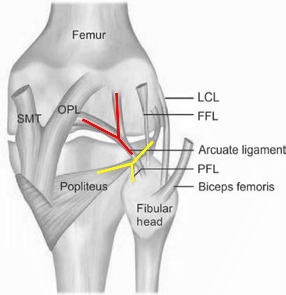
posterolateral corner knee

Biomechanics of PLC

1. Varus restraint: Primary (LCL) and Secondary (Other structures of PLC)

2. Tibial external rotation restraint (esp. at 30-40° flexion): Primary (LCL and popliteus complex) and Secondary (PCL)

Signs and Tests for PLC injury

1. Gait: Varus thrust or Hyperextension varus thrust (apparent tibia vara due to external rotation of tibia on full extension) or walking with slight knee flexion (to avoid instability and stresses on joint and capsule that occurs in hyperextension)

2. Increased external rotation: Dial test

3. External rotation recurvatum test

4. Posterolateral drawer test

5. Reverse pivot shift test

Modified Hughston Classification

GradeI (Mild)II (Moderate)III (Severe)
Varus stress – lateral opening at 30 degrees knee flexion0-5 mm6-10 mm>10 mm
Dial test – rotational instability0-5 degrees6-10 degrees>10 degrees

What is triple varus alignment?

  • Facebook
  • Twitter
PGMEE, MRCS, USMLE, MBBS, MD/MS AnatomyMusculoskeletal systemOrthopedics

Post navigation

Previous post
Next post

Related Posts

PGMEE, MRCS, USMLE, MBBS, MD/MS Advance glaucomatous field defect

Understanding visual field defects in Glaucoma (Perimetry)

May 21, 2014May 21, 2014

Introduction Field of vision or Visual field is defined as the area that is perceived while fixating one central target. According to traquair’s analogy, visual field is “an island of vision surrounded by a sea of darkness”. It is a 3D hill – peak being fovea and at ground level,…

Read More
PGMEE, MRCS, USMLE, MBBS, MD/MS brachial-plexus-block-approaches

Brachial Plexus Block Made Easy

Oct 23, 2016

Brachial plexus is sub-divided from proximal to distal into: Roots, Trunks, Divisions, Cords, Branches. This can be easily remembered with a mnemonic: Randy Travis Drinks Cold Beer. Approaches for Brachial Plexus Block Basically, there are 4 approaches to the brachial plexus block at different levels as described in the mnemonic…

Read More
PGMEE, MRCS, USMLE, MBBS, MD/MS

Internal and External Jugular Veins with Mnemonics

May 18, 2024

Jugular veins are the veins that take blood from the head to the heart via Superior Venacava (SVC). Internal Jugular Vein (IJV) External Jugular Vein (EJV) Formation (Drains from) Inferior petrosal sinus + Sigmoid sinus (At or just distal to Jugular foramen) Posterior div. of retromandibular vein + Posterior auricular…

Read More

Leave a Reply Cancel reply

Your email address will not be published. Required fields are marked *

This site uses Akismet to reduce spam. Learn how your comment data is processed.

Epomedicine. Posterolateral corner (PLC) of knee [Internet]. Epomedicine; 2022 Oct 29 [cited 2026 Jul 14]. Available from: https://epomedicine.com/medical-students/posterolateral-corner-plc-knee/.

Pre-clinical (Basic Sciences)

Anatomy

Biochemistry

Community medicine (PSM)

Embryology

Microbiology

Pathology

Pharmacology

Physiology

Clinical Sciences

Anesthesia

Dermatology

Emergency medicine

Forensic

Internal medicine

Gynecology & Obstetrics

Oncology

Ophthalmology

Orthopedics

Otorhinolaryngology (ENT)

Pediatrics

Psychiatry

Radiology

Surgery

RSS Ask Epomedicine

  • What to study for Clinical examination in Orthopedics?
  • What is the mechanism of AVNRT?

Epomedicine weekly

  • About Epomedicine
  • Contact Us
  • Author Guidelines
  • Submit Article
  • Editorial Board
  • USMLE
  • MRCS
  • Thesis
©2026 Epomedicine | WordPress Theme by SuperbThemes