Skip to content
Epomedicine

Mnemonics, Simplified Concepts & Thoughts

Epomedicine

Mnemonics, Simplified Concepts & Thoughts

Osteochondroma : Mnemonics

Dr. Sulabh Kumar Shrestha, MS Orthopedics, Oct 5, 2022Oct 5, 2022

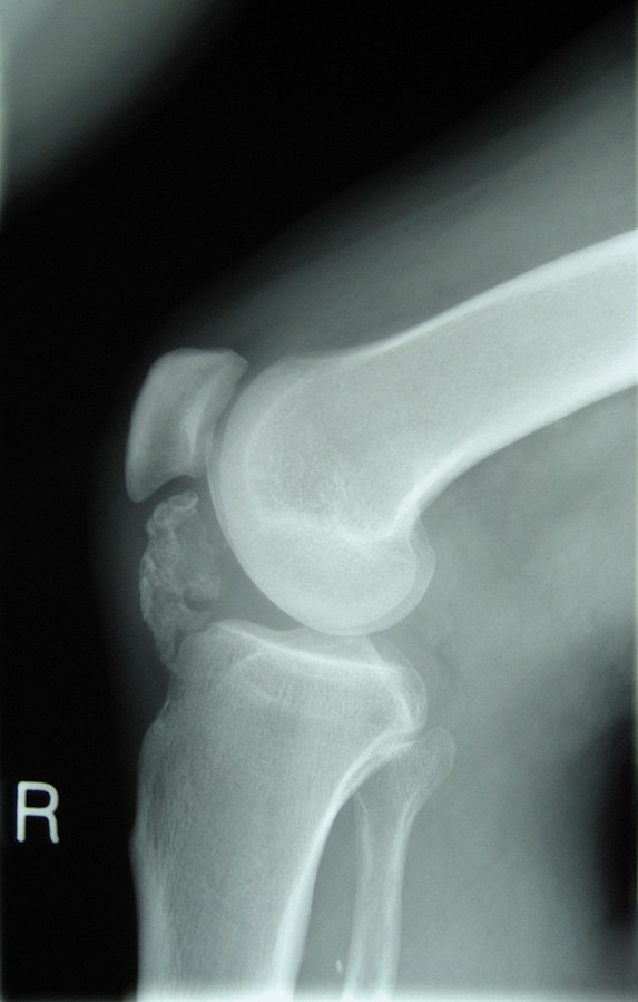
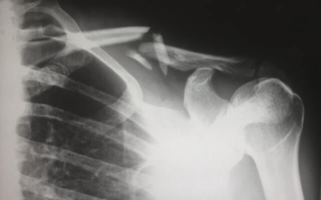
Features of Osteochondroma

Mnemonic: Six “C”

  1. Commonest benign bone tumor
  2. Continue to grow until Closure of physis
  3. Cartilage cap (appears larger clinically than in X-ray)
  4. Continuous with native bone (cortex and medullary canal)
  5. Cane (pedunculated/stalked points away from joint and sessile/broad based have higher risk of malignant degeneration)
  6. Change (mutation) in EXT1, EXT2 and EXT3 genes
osteochondroma
Michael R Carmont, Sian Davies, Daniel Gey van Pittius and Robin Rees, CC BY 2.0, via Wikimedia Commons

Complications

Mnemonic: ABCDE

  1. Adventitious bursa (pain)
  2. Breakage of stalk (pain)
  3. Chondrosarcoma (pain; risk: 1% in solitary osteochondroma and 10% in multiple osteochondroma)
  4. Deformities:
    • Femoral shortening
    • Coxa valga
    • Genu valgum, Ankle valgus (because of shortened fibula)
    • Patellar dislocation
    • Ulnar shortening, radial bowing and ulnar deviation of hand
    • Radial head dislocation
  5. Encroachment:
    • Nerve compression (sciatic, common peroneal, radial)
    • Pseudoaneurysm (popliteal artery)
    • Tendon (rotator cuff impingement, subscapularis tear, bicipital tendinitis)

Common locations

Mnemonic: FGH

  1. Femur (proximal)
  2. Genu (knee)
  3. Humerus (proximal)
  4. Hallux (subungual)

Risk factors for Malignant transformation

Mnemonic: GLAD PaST

  1. Growth after skeletal maturity
  2. Lucency (new)
  3. Additional scintigraphic activity
  4. Destruction (cortical)
  5. Pain after puberty
  6. Soft tissue mass
  7. Thickened cartilage cap >1.5 cm

Pathogenesis theories
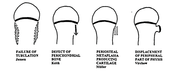
theories osteochondroma

Mnemonic: Five “P”

1. Bone defect

  • Perichondral bone defect and (Keith)

2. Cartilaginous metaplasia

  • Periosteal metaplasia in periphyseal-perimetaphyseal area producing cartilage (Muller)

3. Displacement of physis

  • Peripheral part of physis displacement (Virchow)

4. Epiphysis retains chondrogenic potential

  • Pathological retainment of chondrogenic properties by the epiphysis that would form perichondral bone of the groove of ranvier (Langenskiold)

5. Failure of tubulation

  • Prominent metaphysis suggesting tubulation failure (Jansen)
dr. sulabh kumar shrestha
Dr. Sulabh Kumar Shrestha, MS Orthopedics

He is the section editor of Orthopedics in Epomedicine. He searches for and share simpler ways to make complicated medical topics simple. He also loves writing poetry, listening and playing music. He is currently pursuing Fellowship in Hip, Pelvi-acetabulum and Arthroplasty at B&B Hospital.

  • Facebook
  • Twitter
PGMEE, MRCS, USMLE, MBBS, MD/MS Musculoskeletal systemOncologyOrthopedics

Post navigation

Previous post
Next post

Related Posts

PGMEE, MRCS, USMLE, MBBS, MD/MS internal capsule

Internal Capsule Simplified

Jul 30, 2016

Internal Capsule is a “boomerang” shaped (on horizontal section) and “funnel” shaped, i.e. tapering from superior to inferior (on sagittal section) white matter structure sandwiched between: Medially: Head of Caudate nucleus and Thalamus Laterally: Lenticular nucleus (Globus pallidus and Putamen) Parts of Internal Capsule 1. Anterior limb: Carries fibers to…

Read More
PGMEE, MRCS, USMLE, MBBS, MD/MS Proapoptotic and Antiapoptotic

Apoptosis (Caspases and Genes) : Mnemonics

Jan 5, 2017Aug 13, 2023

Caspases (Pro-apoptotics) Initiator caspases: Executioner caspases: 3 and 7 Pro-apoptotic genes Mnemonic: “B” followed by a vowel Anti-apoptotic genes Mnemonic: Contains “L” or “XL” Other: Inhibitor of Apoptosis (XIAP) How all these genes and molecules work together to complete apoptosis? Find it here.

Read More
PGMEE, MRCS, USMLE, MBBS, MD/MS

Meniscal Tears on MRI

Apr 24, 2025Apr 24, 2025

Meniscal tears are best seen on T1-weighted, gradient-echo and proton-density images. The menisci are low intensity on all sequences. Morphologies Meniscal tear morphology Description MRI appearance Horizontal Separates meniscus into superior (femoral) & inferior (tibial) fragments Primarily horizontal signal on sagittal images Vertical radial Splits central margin of meniscus Vertical…

Read More

Leave a Reply Cancel reply

Your email address will not be published. Required fields are marked *

This site uses Akismet to reduce spam. Learn how your comment data is processed.

Shrestha SK. Osteochondroma : Mnemonics [Internet]. Epomedicine; 2022 Oct 5 [cited 2026 Jul 5]. Available from: https://epomedicine.com/medical-students/osteochondroma-mnemonics/.

Pre-clinical (Basic Sciences)

Anatomy

Biochemistry

Community medicine (PSM)

Embryology

Microbiology

Pathology

Pharmacology

Physiology

Clinical Sciences

Anesthesia

Dermatology

Emergency medicine

Forensic

Internal medicine

Gynecology & Obstetrics

Oncology

Ophthalmology

Orthopedics

Otorhinolaryngology (ENT)

Pediatrics

Psychiatry

Radiology

Surgery

RSS Ask Epomedicine

  • What to study for Clinical examination in Orthopedics?
  • What is the mechanism of AVNRT?

Epomedicine weekly

  • About Epomedicine
  • Contact Us
  • Author Guidelines
  • Submit Article
  • Editorial Board
  • USMLE
  • MRCS
  • Thesis
©2026 Epomedicine | WordPress Theme by SuperbThemes