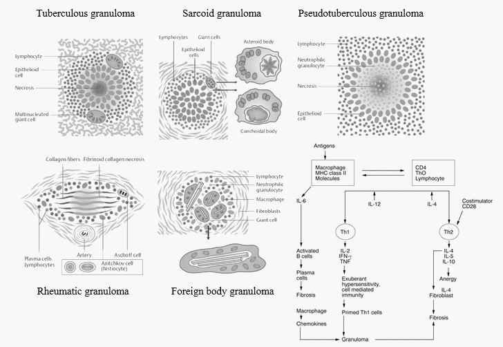
Concentric layers of Granuloma
There are 4 concentric layers in a granuloma, however the clear distinction is difficult in reality due to overlapping. From inside to out:
1. Necrosis
- Caseating necrosis: Tuberculosis, Leprosy
- Coagulative necrosis: Buruli ulcer (M.ulcerans), Gumma containing central blood vessels (Syphilis)
- Fibrinoid necrosis: Aschoff bodies (Rheumatic granuloma), Rheumatoid granuloma, Sarcoid and Beryllium granulomas (rarely)
- Central abscess or microabscess with neutrophils and almost no giant cells (Pseudotuberculous granulomas): Infectious and usually stellate configuration
- Yersinia pseudotuberculosis
- Bartonella (Cat scratch disease)
- Brucellosis
- Tularemia
- Lymphogranuloma venereum
- Listerosis
- Histoplasmosis
- Cryptococcosis
- Typhoid fever
- No necrosis i.e., non-caseating granuloma: Mnemonic – RBCS and LDH
- Reaction to foreign body
- Sarcoid granulomas – Berylliosis, Chron’s disease, Sarcodiosis
- Leprosy – Tuberculoid
- Drug reactions
- Hypersensitivty pneumonitis
- Toxoplasmosis
2. Giant cells, Epitheloid cells and Macrophages
- Epitheloid cells (Macrophages that are secretory and have lost phagocytic properties): Tuberculous granuloma, Sarcoid granuloma, Tumor associated granuloma
- Phagocytic histiocytes: Foreign body granuloma, Rheumatic and Rheumatoid granuloma
- Langhans giant cells (Horse shoe pattern of nuclear arrangement): Tuberculous granuloma (Tuberculosis, Leprosy, Syphilis)
- Foreign body giant cells (Haphazard and random nuclear arrangement): Foreign bodies like suture, talc, etc.
- Touton giant cells (Ring of nuclei surrounded by foamy pale cytoplasm): Xanthoma, Fat necrosis, Xanthogranulomatous inflammation, Dermatofibroma
- Palisading granuloma (Elongated nuceli of mononuclear phagocytes are palisaded): Rheumatoid nodules, Post-surgical necrobiotic granuloma in prostate/urinary bladder, etc.
- Asteroid bodies (stellate inclusions with numerous rays radiating from a central core) and Schaumann or conchoidal bodies (calcium deposited asteroid bodies): Sarcoid granuloma (Hard sore)
- No giant cells: Pseudotuberculous granuloma
3. Lymphocytes
- Especially prominent in immune granulomas.
- Lymphocytes secrete mediators that activate and alter macrophages and macrophage-derived cells located centrally.
4. Fibroblasts
- Walls off the lesion.
Pathophysiology and Morphology of Granuloma