Median nerve, Ulnar nerve and Radial nerve are the 3 major nerves of the upper limb originating from the brachial plexus.
Some important neuroanatomic relationships in the forearm are:
1. Medial nerve: Crossed by brachial artery from lateral to medial just above the elbow to lie medial to brachial artery in cubital fossa.
2. Anterior interosseous nerve: Splits pronator teres and runs between flexor digitorum superficialis (FDS) and flexor digitorum profundus (FDP).
3. Ulnar nerve: Between flexor carpi ulnaris (FCU) and flexor digitorum profundus (FDP).
4. Radial nerve: Between brachialis and brachioradialis.
5. Posterior interosseous nerve: Splits supinator
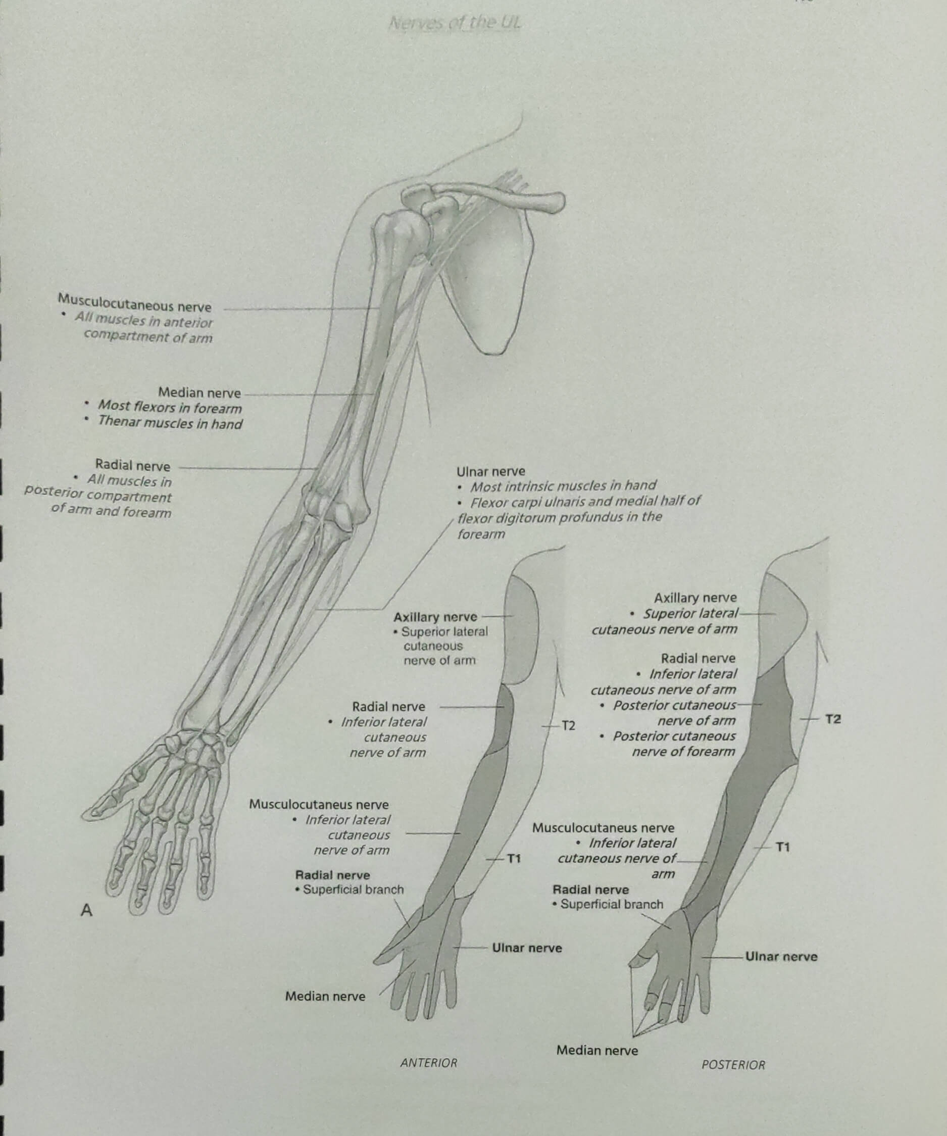
Course and innervation of these nerves have been simplified and tabulated below:
| Part of upper limb | Ulnar nerve | Supply | Median nerve | Supply | Radial nerve | Supply |
| Origin | Medial cord (C8-T1) | Lateral and Medial cord (C5-T1) | Posterior cord (C5-T1) | |||
| Axilla | Medial to axillary artery | None | Lateral to axillary artery | None | Behind axillary artery – exits axilla via Triangular interval | Long and medial head of triceps Posterior cutaneous nerve of arm |
| Proximal arm | Medial to brachial artery | None | Lateral to brachial artery | None | Posterior compartment (Spiral groove – from medial to lateral) | Lateral head of triceps and Anconeus Lateral cutaneous nerve of arm and Posterior cutaneous nerve of forearm |
| Distal arm | Posterior compartment (over medial head of triceps) after piercing medial intermuscular septum | None | Anterior compartment (Crosses brachial artery from lateral to medial at coracobrachialis insertion) – runs under biceps | None | Anterior compartment (Pierces lateral intermuscular septum ~10 cm proximal to lateral epicondyle) – in front of lateral epicondyle between Brachialis and BR/ECRL | Brachialis BR ECRL |
| Elbow | Passes behind medial epicondyle (cubital tunnel) | Elbow joint | Cubital fossa (Medial to brachial artery; Beneath lacertus fibrosus) | Elbow joint | Divides | |
| Forearm | Enters between 2 heads of FCU | FCU | Enters between 2 heads of PT | PT | SBRN – sensory (travels under BR and emerges between BR and ECRL ~8 cm proximal to radial styloid) | |
| Travels between FCU and FDP | Ulnar 2 FDPs | Branches to anterior compartment muscles of forearm | PL, FCR, FDS | DBRN (passes through FREAS) -Fibrous bands – Radial neck (radial recurrent vessels) – ECRB – Arcade of Frohse (supinator arch) -Supinator (distal border) | ||
| Palmar cutaneous branch | Hypothenar skin | Anterior interosseous nerve – runs between FDP and FPL | PQ, FPL, lateral 2 FDP | DBRN emerges out of Radial tunnel/FREAS as PIN | Supinator and all 6 muscles of extensor compartment | |
| Dorsal cutaneous branch | Dorsal aspect of medial 1 & 1/2 fingers and associated hand area | Travels down forearm entering 2 heads of FDS (sublimis bridge) and runs between FDS and FDP | ||||
| Runs between FCR and PL (gives palmar cutaneous branch) | Thenar eminence sensory | |||||
| Wrist and Hand | Passes superficial to flexor retinaculum and travels under piso-hamate ligament (Guyon’s canal) | Passes deep to carpal tunnel (flexor retinaculum) | SBRN runs as dorsal radial sensory nerve crossing anatomical snuff box lateral to radial artery | Wrist, Dorsal lateral 3 and 1/2 fingers upto distal interphalangeal joint and corresponding dorsum of hand | ||
| a. | Superficial branch (sensory) | Palmar surface of medial 1 & 1/2 fingers Palmaris brevis | Palmar digital branch | Palmar surface and fingertips of lateral 3 and 1/2 digits 1st and 2nd lumbricals | ||
| b. | Deep branch (motor) | 1 & 1/2 thenar muscles – Adductor pollicis, deep head of FPB All hypothenar muscles except palmaris brevis All interossei 2 Lumbricals (3rd, 4th) | Recurrent thenar branch | Thenar muscles (AbPB, OP, Superficial head of FPB) |
Further reading: