Origin
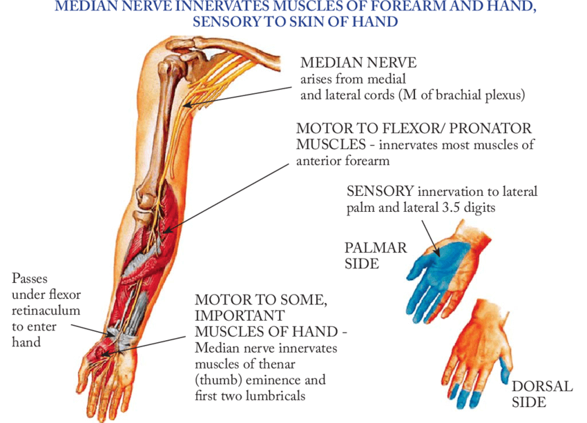
C5-T1 (lateral and medial cords of Brachial plexus)
Course
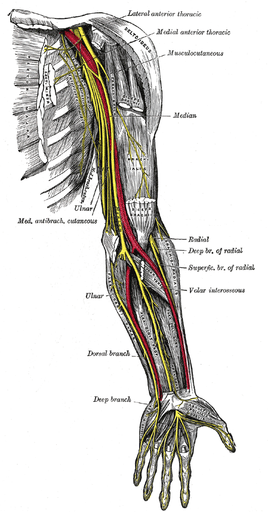
- Axilla: Starts in axilla (lateral or anterior to axillary artery)
- Arm: Runs along with brachial artery – initially lateral to it (in arm) and later medial to it (cubital fossa); i.e. crosses brachial artery from lateral to medial on mid-arm
- Anatomical variations: lateral and medial cords may fuse at as low as elbow; may receive lateral cord contribution through musculocutaneous nerve
- Antecubital fossa/Elbow: Enters antecubital fossa passing medial to biceps and over the brachialis; Passes through 3 successive arches to enter forearm
- Bicipital aponeurosis (Lacertus fibrosus)
- 2 heads of pronator teres (superficial humeral and deep ulnar heads; may have only 1 head as anatomical variation)
- 2 heads of FDS or sublimis (medial humeroulnar head and lateral radial head; may have only 1 head as anatomical variation) under the sublimis ridge
- Forearm: Travels between FDS (superficial) and FDP (deep)
- Gives Anterior Interosseous Nerve (AIN) branch which travels on interosseous membrane between and below FDP and FPL and terminates in the distal forearm deep to PQ
- Nerve continues down to become superficial about 5 cm proximal to wrist crease, between FCR (laterally) and Palmaris longus tendon (medially) and gives Palmar cutaneous branch (runs superficial to carpal tunnel)
- Wrist: Passes under the carpal tunnel
- Hand:
- Recurrent thenar motor branch (Variations: can originate in carpal tunnel; can pierce transverse carpal ligament)
- Digital branches (superficial to flexor tendons but deep to superficial palmar arterial arch)
Motor innervation
Muscles supplied: 2 lateral lumbricals, 3 thenars, 3Ps (2 pronator and 1 palmaris) and 4 flexors.
- Superficial forearm: Pronator teres, Flexor carpi radialis, Palmaris longus (All superficial muscles except flexor carpi ulnaris)
- Intermediate forearm: Flexor digitorum superficialis
- Deep forearm: Flexor digitorum profundus (medial half supplied by ulnar nerve), Flexor pollicis longus, Pronator quadratus (by Anterior Interosseous Nerve or AIN)
- Hand: 1st and 2nd lumbricals, Oppones pollicis, Abductor pollicis brevis, Flexor pollicis brevis (All thenar muscles except adductor pollicis longus which is supplied by ulnar nerve)
Sensory innervation
- Palmar cutaneous branch: Lateral palm
- Digital cutaneous branch: Lateral 3 and 1/2 digits and palm on the palmar side and only fingertips on dorsal side
Clinical correlation
1. Martin-Gruber motor connection: occur in 17% of individuals between median and ulnar nerves resulting in variable innervations of intrinsic muscles.
2. Pronator syndrome: It presents with pain on volar aspect of distal arm and proximal forearm which may be increased by flexion of elbow or middle finger (FDS) against resistance. Entrapment neuropathy of median nerve in elbow; at 4 sites:
- Ligament of struthers (connecting supracondylar spur to accessory origin of pronator teres)
- Bicipital aponeurosis
- Aponeurotic edges of deep head of pronator teres
- Tendinous aponeurotic arch forming proximal free edge of radial attachment of Flexor Digitorum Superficialis (FDS)
3. Anterior interosseous nerve palsy: Weakness of pinch grip (due to involvement of flexor pollicis longus and flexor digitorum profundus of index finger) and no sensory symptoms (differentiating feature from pronator syndrome)
4. Injury:
- At wrist level: Positive pen test (paralysis of abductor pollicis brevis) – unable to touch the object held above the thumb at right angle to palm and Ape thumb deformity (paralysis of abductor pollicis brevis) – adducted and laterally rotated thumb + Loss of opposition and abduction of thumb.
- At elbow level: Above + Supinated forearm (pronators paralyzed) + Weak wrist flexion (Paralyzed long flexors except FCU and medial 1/2 FDP) + Adducted wrist (Paralyzed FCR and Intact FCU) + Loss of flexion of terminal phalanx of thumb (FPL paralyzed) + Pointing index/Positive Oschner clasp/Benediction test (Paralysis of FDS and lateral 1/2 FDP = flexion of interphalangeal joint of index and middle fingers is lost)
CONFUSED !!