Skip to content
Epomedicine

Mnemonics, Simplified Concepts & Thoughts

Epomedicine

Mnemonics, Simplified Concepts & Thoughts

Kanavel Sign for Pyogenic Flexor Tenosynovitis

Epomedicine, Sep 30, 2020Sep 30, 2020

1. Exquisite tenderness over the course of the sheath, limited to the sheath

  • Present in 64% cases
  • Late sign of proximal extension of pyogenic tenosynovitis
  • Most important sign as described by Kanavel

2. Flexion of the finger (‘hook’ sign)

  • Present in 69% cases

3. Exquisite pain on extending the finger, most marked at the proximal end

  • Present in 72% cases
  • Most reliable early Kanavel sign

4. Fusiform swelling of the digit (‘sausage’ digit)

  • Present in 97% cases

About only 54% patients have all 4 signs.

Kanavel signs

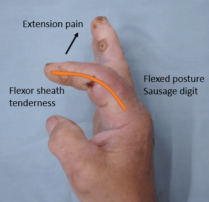
Additional sign by Neviaser and Gunther: inability to flex the finger to touch the palm 

Kanavel’s signs in the little finger and thumb may be more subtle than the central fingers because of autodecompression through the ulnar and radial bursae.

Reference: Kennedy CD, Huang JI, Hanel DP. In Brief: Kanavel’s Signs and Pyogenic Flexor Tenosynovitis. Clin Orthop Relat Res. 2016 Jan;474(1):280-4. doi: 10.1007/s11999-015-4367-x. Epub 2015 May 29. PMID: 26022113; PMCID: PMC4686527.

  • Facebook
  • Twitter
PGMEE, MRCS, USMLE, MBBS, MD/MS Musculoskeletal systemOrthopedics

Post navigation

Previous post
Next post

Related Posts

PGMEE, MRCS, USMLE, MBBS, MD/MS calots triangle

Calot’s triangle : Mnemonic

Mar 2, 2018Aug 17, 2023

Synonyms: Calot triangle, Cystohepatic triangle Boundaries: Cysto-hepatic triangle (Budde-Rocko triangle): Calot’s triangle as described in modern surgery: Mnemonic: 3 C 1. Cystic duct 2. Common hepatic duct 3. Cystic artery Contents: Importance: The cystic artery and the duct have to be clearly defined to obtain the ‘critical view of safety’. These…

Read More

Anatomy and Physiology of Sebaceous glands

Nov 11, 2013

Definition: Sebaceous glands are numerous microscopic glands in the dermis that usually open into the hair follicles and secrete sebum. They are holocrine glands, i.e., the sebum consists of the entire secreting cells. Location: Found everywhere on the skin apart from the palms and soles Types of Sebaceous glands: 1….

Read More
PGMEE, MRCS, USMLE, MBBS, MD/MS

Ollier’s Disease and Maffucci Syndrome : Mnemonic

Dec 1, 2024Dec 1, 2024

Ollier’s disease Maffucci syndrome

Read More

Leave a Reply Cancel reply

Your email address will not be published. Required fields are marked *

This site uses Akismet to reduce spam. Learn how your comment data is processed.

Epomedicine. Kanavel Sign for Pyogenic Flexor Tenosynovitis [Internet]. Epomedicine; 2020 Sep 30 [cited 2026 Jun 27]. Available from: https://epomedicine.com/medical-students/kanavel-sign-pyogenic-flexor-tenosynovitis/.

Pre-clinical (Basic Sciences)

Anatomy

Biochemistry

Community medicine (PSM)

Embryology

Microbiology

Pathology

Pharmacology

Physiology

Clinical Sciences

Anesthesia

Dermatology

Emergency medicine

Forensic

Internal medicine

Gynecology & Obstetrics

Oncology

Ophthalmology

Orthopedics

Otorhinolaryngology (ENT)

Pediatrics

Psychiatry

Radiology

Surgery

RSS Ask Epomedicine

  • What to study for Clinical examination in Orthopedics?
  • What is the mechanism of AVNRT?

Epomedicine weekly

  • About Epomedicine
  • Contact Us
  • Author Guidelines
  • Submit Article
  • Editorial Board
  • USMLE
  • MRCS
  • Thesis
©2026 Epomedicine | WordPress Theme by SuperbThemes