Skip to content
Epomedicine

Mnemonics, Simplified Concepts & Thoughts

Epomedicine

Mnemonics, Simplified Concepts & Thoughts

voulantary horizontal conjugate gaze

Horizontal Conjugate Gaze Pathway

Epomedicine, Jul 27, 2016May 19, 2019

Components of Pathway

For both eyes to look at a side:

  1. Contralateral Frontal Eye Field (Brodmann area 8)
  2. Ipsilateral PPRF (Paramedial Pontine Reticular Formation)
  3. Ipsilateral CN VI Nucleus
  4. Contralateral Medial Longitudinal Fasciculus (MLF)
  5. Contralateral CN III Nucleus

Horizontal Conjugate Gaze Pathway

voulantary horizontal conjugate gaze

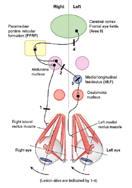
Lesions of Conjugate Gaze Pathway

Abducens (CN VI) nerve:

Affected side cannot abduct

Lesion site 1 in figure – Right abducens nerve

  • Right eye cannot look right

Abducens (CN VI) nucleus or PPRF:

Both the eyes cannot look towards the affected side (Lateral gaze paralysis)

Lesion site 2 in figure – Right abducens nucleus

  • Right eye cannot look right or abduct (Right Lateral rectus dysfunction)
  • Left eye cannot look right or adduct (Left Medial rectus dysfunction – as abducens nucleus activates contralateral CN III through contralateral MLF)

Medial Longitudinal Fasciculus (MLF):

Eye on affected side cannot adduct when the eye on unaffected side abducts (Internuclear ophthalmoplegia)

Lesion site 3 in figure – Left MLF

  • Left eye cannot adduct or look right when right eye looks to the right
  • Convergence intact
  • Right eye exhibits nystagmus

Can be differentiated from the Occulomotor nerve lesion by the absence of ptosis and mydriasis seen with occulomotor nerve lesion.

Internuclear ophthalmoplegia: Lack of communication such that when CN VI nucleus activates ipsilateral lateral rectus, contralateral CN III nucleus does not stimulate medial rectus to contract. Abducting eye displays nystagmus (CN VI overfires to stimulate CN III). Convergence is normal.

If the left eye is able to abduct and shows nystagmus, the pathway involves –

  1. Ipsilateral (left) structures – Pontine structures (PPRF and CN VI)
  2. Contralateral (right) structures – Structures above pons (MLF, CN III and frontal eye field)
  3. Diagnosis – Right Intranuclear ophthalmoplegia (INO) because right MLF is involved

Mnemonic:

  1. INO – Impaired adduction and Nystagmus Opposite (i.e. Left INO = Left impaired adduction and Right nystagmus)

Frontal Eyefield:

Eyes cannot look to the side opposite to the lesion

Lesion site 4 in figure – Left Frontal eye field

  • Both the eyes cannot look to the right
  • Slowly drift to the left
13 shares
  • Facebook8
  • Twitter
PGMEE, MRCS, USMLE, MBBS, MD/MS AnatomyNervous systemOphthalmology

Post navigation

Previous post
Next post

Related Posts

PGMEE, MRCS, USMLE, MBBS, MD/MS calots triangle

Calot’s triangle : Mnemonic

Mar 2, 2018Aug 17, 2023

Synonyms: Calot triangle, Cystohepatic triangle Boundaries: Cysto-hepatic triangle (Budde-Rocko triangle): Calot’s triangle as described in modern surgery: Mnemonic: 3 C 1. Cystic duct 2. Common hepatic duct 3. Cystic artery Contents: Importance: The cystic artery and the duct have to be clearly defined to obtain the ‘critical view of safety’. These…

Read More
PGMEE, MRCS, USMLE, MBBS, MD/MS gamma loop

Upper Motor Neuron Lesions (UMNL) – Anatomical Basis

Jul 24, 2016Dec 7, 2022

For the purpose of remembering the clinical manifestations of upper motor neuron lesion (UMNL) and lower motor neuron lesion (LMNL), a mnemonic has already been devised and discussed here. Now, it’s time to understand the anatomical and physiological basis of these manifestations. Upper Motor Neuron Lesion (UMNL) Syndrome Acute Manifestations…

Read More
PGMEE, MRCS, USMLE, MBBS, MD/MS

Thyroid Hormone Function & Synthesis : Mnemonics

Aug 19, 2023May 21, 2024

Thyroid Hormone Functions Mnemonic: 7 X B’s Thyroid Hormone Synthesis Mnemonic: ATE ICE 1. Active transport of iodide: 2. Thyroglobulin (rich in Tyrosine): formed in follicular ribosomes 3. Exocytosis of thyroglobulin into follicular lumen: stored as colloid 4. Iodination: Peroxidase enzyme – 5. Coupling: 6. Endocytosis: of T3 and T4…

Read More

Comments (2)

  1. Kokki says:
    Feb 14, 2020 at 6:55 am

    Concept cleared! It has been so confusing like forever!

    Reply
  2. Asmita Paudel says:
    Oct 8, 2021 at 10:27 am

    Thank you so much. I never knew this concept is this easy. Hats off!

    Reply

Leave a Reply to Kokki Cancel reply

Your email address will not be published. Required fields are marked *

This site uses Akismet to reduce spam. Learn how your comment data is processed.

Pre-clinical (Basic Sciences)

Anatomy

Biochemistry

Community medicine (PSM)

Embryology

Microbiology

Pathology

Pharmacology

Physiology

Clinical Sciences

Anesthesia

Dermatology

Emergency medicine

Forensic

Internal medicine

Gynecology & Obstetrics

Oncology

Ophthalmology

Orthopedics

Otorhinolaryngology (ENT)

Pediatrics

Psychiatry

Radiology

Surgery

RSS Ask Epomedicine

  • What to study for Clinical examination in Orthopedics?
  • What is the mechanism of AVNRT?

Epomedicine weekly

  • About Epomedicine
  • Contact Us
  • Author Guidelines
  • Submit Article
  • Editorial Board
  • USMLE
  • MRCS
  • Thesis
©2026 Epomedicine | WordPress Theme by SuperbThemes