Skip to content
Epomedicine

Mnemonics, Simplified Concepts & Thoughts

Epomedicine

Mnemonics, Simplified Concepts & Thoughts

gastric glands

Gastric glands and cells

Epomedicine, Jul 15, 2018Jun 16, 2023

Anatomically, stomach can be divided into 3 parts from above to below:

  1. Cardiac
  2. Fundus and body/corpus
  3. Pyloric/Antral

Gastric pits deepen as we move below from cardiac to pyloric glands.

Mucus secreting cells: Present in all 3 glands – cardiac, fundic and pyloric but predominate in cardiac and pyloric glands.

Pyloric glands have 1 more cell – G cells which secrete Gastrin.

gastric glands

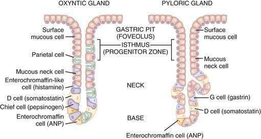
Fundic glands (gastric glands of fundus and body) can be divided into 3 parts from above (towards gastric pit) to downwards:

  1. Isthmus (Outer zone) – Mucus cells and Stem cells
  2. Neck (Middle zone) – Parietal (Oxyntic) cells
  3. Fundus (Basal zone) – Chief (Zymogen) cells and Enteroendocrine cells (Mnemonic: ECF)

Parietal (Oxyntic) cells secrete:

  1. Hydrochloric acid
  2. Intrinsic factor
  3. Ghrelin

Mnemonic: Parietal cells are Pink (Eosinophilic).

Chief (Zymogen/Peptic) cells secrete:

  1. Pepsin
  2. Gastric lipase

Mnemonic: Basal cheif cells are Blue (Basophilic). These have a lot of rough endoplasmic reticulum (rER).

Enteroendocrine cells:

  • G cells = Gastrin
  • D cells = Somatostatin
  • ECL cells = Histamine
6 shares
  • Facebook6
  • Twitter
PGMEE, MRCS, USMLE, MBBS, MD/MS AnatomyGastrointestinal systemPhysiology

Post navigation

Previous post
Next post

Related Posts

PGMEE, MRCS, USMLE, MBBS, MD/MS chemokines

Chemokines Mnemonics

Jan 18, 2017Jan 18, 2017

Difference between cytokines and chemokines Cytokines are small proteins released by cells, the function of which is “cell-signaling“. Chemokines are small cytokines, which functions as a “chemo-attractant“. Types of Chemokines When you go through the structural classification of chemokines, you come accross various arrangements of letter: C: denotes cysteine X:…

Read More
PGMEE, MRCS, USMLE, MBBS, MD/MS rib notching

Rib Notching

Feb 13, 2017

Normal Rib Notching A small notch near the costo-vertebral joint is normal, so pathologic rib notching is more likely if the notching is more lateral. Types of Pathological Rib Notching 1. Superior rib notching 2. Inferior rib notching (more common) Inferior Rib Notching (Roesler’s sign) Mechanism: Enlargement of one or…

Read More
PGMEE, MRCS, USMLE, MBBS, MD/MS opioid receptors

Opioid Adverse Effects – Mnemonic

Apr 14, 2021Apr 14, 2021

Mnemonic: MORPHINES 1. Miosis Mechanism: Stimulation of Edinger-Westphal nucleus of Cranial nerve III 2. Orthostatic hypotension Mechanism: Vasodilation due to – Direct action decreasing tone of blood vessels Histamine release Depression of vasomotor center 3. Respiratory depression Mechanism: Inhibits respiratory center in a dose-dependent manner (neurogenic, hypercapneic and hypoxic drives…

Read More

Leave a Reply Cancel reply

Your email address will not be published. Required fields are marked *

This site uses Akismet to reduce spam. Learn how your comment data is processed.

Pre-clinical (Basic Sciences)

Anatomy

Biochemistry

Community medicine (PSM)

Embryology

Microbiology

Pathology

Pharmacology

Physiology

Clinical Sciences

Anesthesia

Dermatology

Emergency medicine

Forensic

Internal medicine

Gynecology & Obstetrics

Oncology

Ophthalmology

Orthopedics

Otorhinolaryngology (ENT)

Pediatrics

Psychiatry

Radiology

Surgery

RSS Ask Epomedicine

  • What to study for Clinical examination in Orthopedics?
  • What is the mechanism of AVNRT?

Epomedicine weekly

  • About Epomedicine
  • Contact Us
  • Author Guidelines
  • Submit Article
  • Editorial Board
  • USMLE
  • MRCS
  • Thesis
©2026 Epomedicine | WordPress Theme by SuperbThemes