Skip to content
Epomedicine

Mnemonics, Simplified Concepts & Thoughts

Epomedicine

Mnemonics, Simplified Concepts & Thoughts

Deltoid Ligament : Anatomy, Clinical tests and Acute Injury

Dr. Sulabh Kumar Shrestha, MS Orthopedics, Oct 23, 2024Oct 23, 2024

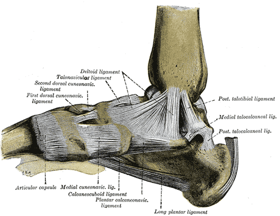
Deltoid ligament is the complex of medial collateral ligaments of the ankle joint.

2 layers of Deltoid Ligament

a. Superficial layer: Originates from anterior and inferior aspects of medial malleolus fanning out & sending 3 bands to –

  • Navicular (Tibionavicular ligament) and Plantar calcaneonavicular or spring ligament (Tibiospring ligament)
  • Sustenaculum tali of calcaneus (Tibiocalcaneal ligament)
  • Medial tubercle of talus (Superficial posterior tibiotalar ligament)

b. Deep layer: Originates on posterior border of anterior colliculus, intercollicular groove & posterior colliculus and inserts into entire non-articular surface of medial talus. It is intra-articular and covered by synovium and comprises of 2 ligaments –

  • Anterio tibiotalar ligament
  • Deep posterior tibiotalar ligament
deltoid ligament
Henry Vandyke Carter, Public domain, via Wikimedia Commons

Clinical tests for Deltoid Ligament

1. Eversion test in neutral: Evaluates superficial deltoid ligament

2. External rotation stress test: Evaluates syndesmotic ligaments + deep deltoid ligament

Acute Deltoid Ligament Injury

Mechanism of injury:

  1. Eversion and/or pronation
  2. Associated with lateral ankle fractures
Ligament Tests for Ankle Injuries

Hintermann’s classification:

  • Type I – Proximal tear or avulsion (70%)
  • Type II – Intermediate tear, i.e. deep component remains attached distally; superficial component remains attached proximally (10%)
  • Type III – Distal tear or avulsion of deltoid & spring ligaments (20%)

Clinical finding:

  • Medial ecchymosis appears after 24 hours
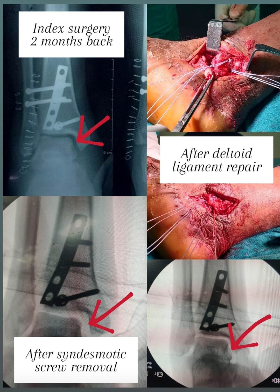
X-ray findings:

  1. Small flake of medial malleolus may be visible (avulsion from tibial attachment)
  2. Lateral talar shift and widened medial clear space between talus and medial malleolus (>5 mm on external rotation stress or gravity stress views)

Treatment:

The deltoid ligament usually heals after functional treatment of the ankle sprain, syndesmotic screw placement, or fixation of the fibula fracture. As talus follows fibula when deltoid is ruptured, anatomic restoration of fibula and talus restores medial anatomy & allows medial ligamentous structures to heal without the need for operative treatment.

deltoid ligament repair using suture anchors
Red arrow indicating widened medial clear space before treatment and reduced medial clear space after deltoid ligament repair with suture anchors.

Surgical repair of the deltoid ligament is indicated only in the rare case of torn ligament blocking reduction of the medial ankle joint. Reduction may be blocked by interposed fibers of deltoid ligament or interposed posterior tibial tendon. Evidence for mal-reduction includes medial clear space is widened by > 2 mm following reduction.

dr. sulabh kumar shrestha
Dr. Sulabh Kumar Shrestha, MS Orthopedics

He is the section editor of Orthopedics in Epomedicine. He searches for and share simpler ways to make complicated medical topics simple. He also loves writing poetry, listening and playing music. He is currently pursuing Fellowship in Hip, Pelvi-acetabulum and Arthroplasty at B&B Hospital.

1 shares
  • Facebook
  • Twitter
PGMEE, MRCS, USMLE, MBBS, MD/MS Musculoskeletal systemOrthopedics

Post navigation

Previous post
Next post

Related Posts

PGMEE, MRCS, USMLE, MBBS, MD/MS

Dermatomes : Mnemonic

Sep 18, 2021Sep 18, 2021

Note: C1 doesn’t have a sensory branch For the purpose of making learning easier, we divide the remaining dermatomal segments into 4 groups which will be accompanied by analogies and mnemonics. C2-C4 These supply the “hooded cape” region. C2: hood region abutting the trigeminal distribution C3: upper neck C4: lower…

Read More
PGMEE, MRCS, USMLE, MBBS, MD/MS hematopoiesis

Hematopoietic growth factors list with mnemonics

May 23, 2019May 23, 2019

a. SCF (Steel factor): Multipontent stem cells b. IL-3: Myeloid stem cells (“3” turned down looks like “m“) c. IL-7: Lymphoid stem cell (“L” turned up looks like “7“) d. GM-CSF (Granulocyte macrophage Colony stimulating factor): Myeloid lineage (Erythrocytes, Thrombocytes, Granulocytes and Monocytes) Pharmaceutical: Sargramostim e. G-CSF: Granulocytes (Neutrophil, Eosinophil,…

Read More
PGMEE, MRCS, USMLE, MBBS, MD/MS

Cervical Vertebrae Anatomy

May 4, 2024May 4, 2024

Osteology Vertebra Body Spinous process Transverse process Typical (C3-6) Relatively small; oval; uncinate process superiorly Bifid Has foramen transversarium, Has anterior and posterior tubercle separated by groove for spinal nerve (large anterior tubercle in C6 called Carotid/Chassaignac tubercle) Atlas (C1) None; Ring (2 lateral masses connected by anterior & posterior…

Read More

Leave a Reply Cancel reply

Your email address will not be published. Required fields are marked *

This site uses Akismet to reduce spam. Learn how your comment data is processed.

Shrestha SK. Deltoid Ligament : Anatomy, Clinical tests and Acute Injury [Internet]. Epomedicine; 2024 Oct 23 [cited 2026 Jun 23]. Available from: https://epomedicine.com/medical-students/deltoid-ligament-anatomy-clinical-tests-and-acute-injury/.

Pre-clinical (Basic Sciences)

Anatomy

Biochemistry

Community medicine (PSM)

Embryology

Microbiology

Pathology

Pharmacology

Physiology

Clinical Sciences

Anesthesia

Dermatology

Emergency medicine

Forensic

Internal medicine

Gynecology & Obstetrics

Oncology

Ophthalmology

Orthopedics

Otorhinolaryngology (ENT)

Pediatrics

Psychiatry

Radiology

Surgery

RSS Ask Epomedicine

  • What to study for Clinical examination in Orthopedics?
  • What is the mechanism of AVNRT?

Epomedicine weekly

  • About Epomedicine
  • Contact Us
  • Author Guidelines
  • Submit Article
  • Editorial Board
  • USMLE
  • MRCS
  • Thesis
©2026 Epomedicine | WordPress Theme by SuperbThemes