Skip to content
Epomedicine

Mnemonics, Simplified Concepts & Thoughts

Epomedicine

Mnemonics, Simplified Concepts & Thoughts

myeloid differentiation aml

Concept of Acute Myeloid Leukemia (AML) FAB Classification

Epomedicine, Aug 23, 2016Mar 10, 2018

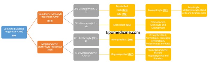
There is no need of mnemonics to remember the FAB classification of Acute Myeloid Leukemia (AML); just remember the process myeloid differentiation. A simple schematic diagram with few intermediate processes and stimulating factors eliminated will meet our purpose here.
myeloid differentiation aml

The cells belonging to the myeloid lineage are:

  1. Granulocytes: Neutrophils, Eosinophils and Basophils
  2. Monocytes and Macrophages
  3. Erythrocytes (RBCs)
  4. Megakaryocytes (Platelets)

In French-American-British (FAB) classification of AML, it is classified from M0 to M7. The scheme takes into account:

  • The degree of maturation (M0 to M3)
  • The lineage of leukemic blast (M4 to M7)
  1. M0 – undifferentiated progenitor cells
  2. M1 to M3 – myelocytes (granulocyte precursors)
  3. M4 to M5 – monocyte precursors
  4. M6 – erythrocyte precursors
  5. M7 – platelet precursors

Simplified FAB classification of AML

  1. M0 – Undifferentiated
  2. M1 – Myeloblastic without maturation
  3. M2 – Myeloblastic with maturation (Commonest type)
  4. M3 – Promyelocytic
  5. M4 – Myelomonocytic (Naegeli type)
  6. M5 – Monocytic (Schilling type)
  7. M6 – Erythroleukemia (Di Gulielmo’s disease)
  8. M7 – Megakaryocytic

AML Concepts in Concise

1. FAB used 30% blasts to delineate chronic myeloid leukemia (CML) from Blast crisis and AML. WHO revised classification uses the presence of ≥20% myeloblasts in the bone marrow or peripheral blood for the diagnosis of AML.

2. Mo, M1 and M2:

  • <3% blasts are MPO positive in M0 and ≥3% blasts are MPO positive in M1
  • <10% maturation beyond myeloblasts = M1
  • >10% mauration beyond myeloblasts = M2
  • Auer rods:
    • M0 – None
    • M1 – 50%
    • M2 – 70%
    • M3 – 100%
  • t(8:21) is pressent in M2

3. M3:

  • Most cases have t(15:17) – results in disruption of Retinoic Acid Receptor (RAR) required for myeloblast maturation.
  • All-trans-retinoic acid is hence, used for treatment of Acute Promyelocytic Leukemia (APL).
  • DIC is seen in 5-10% cases due to prothrombotic release of Auer rods.

4. M4: >20% monocytic precursors (<20% in M2)

  • Myeloperoxidase positive (Myeloblastic) + Auer rod positive (Monoblastic) + Non-specific esterase positive (Monoblastic)

5. M5:

  • >80% monocytic precursors
  • Gum infiltration and hyperplasia
  • High lysozyme level
  • Auer rod negative + Non-specific esterase positive

6. M6:

  • ≥50% erythroid precursors and ≥20% blasts in nonerythroid component
  • Also known as Di Gulielmo’s disease or Syndrome

7. M7:

  • Rapid myelofibrosis due to release of PDGF.
  • Resistant to treatment
  • GATA1 mutations is seen in those associated with Down’s Syndrome.
  • GpIIb/IIIa or vWF positive

8. Ara-C (Cytarabine) is used for the treatment of CML except M3, in which all-trans retinoic acid is used.

9. Remission criteria:

  • Complete remission = Bone marrow blasts <5%, No auer rods
  • Complete blood count recovery = ANC >1000/microlitre and Platelets ≥1,00,000/microlitre
  • Partial remission = ≥50% fall in blasts over pretreatment values

10. M8:

  • There is no M8 in FAB classification, but some authors have proposed rare deno-vo Acute Basophilic Leukemia as M8.
  • Facebook
  • Twitter
PGMEE, MRCS, USMLE, MBBS, MD/MS HematologyInternal medicinePathology

Post navigation

Previous post
Next post

Related Posts

PGMEE, MRCS, USMLE, MBBS, MD/MS retinal layers

Retinal Layers Simplified

Jul 28, 2016Oct 29, 2017

The ten layers of retina – this microscopic anatomy is frequently asked in examinations and also important from the physiological viewpoint. There are plenty of mnemonics around the web, but we will proceed in a different approach to remember the 10 retinal layers easily. A. Retina is 3 neuron system…

Read More
PGMEE, MRCS, USMLE, MBBS, MD/MS glucose-6-phosphate

Glucose 6 Phosphate : Central to Glucose Metabolism

Jan 22, 2017

Glucose-6-Phosphate central to the 4 major metabolic pathways of glucose, i.e. glycolysis, gluconeogenesis, glycogenesis, glycogenolysis and HMP shunt (Pentose phosphate pathway). Glucose is immediately phosphorylated inside the cells to Glucose-6-Phsophate to trap them inside cell and prevent diffusion out of the cell. Glucose-6-Phosphate is the key intermediate to understand the…

Read More
PGMEE, MRCS, USMLE, MBBS, MD/MS dependence cycle

Substance abuse vs Substance dependence

Nov 4, 2016Nov 10, 2016

Both of these substance use disorders are maladpative patterns of substance use, leading to clinically significant impairment/distress but defined by separate criteria. Substance abuse ≥1 of the following occurring within a 12 month period: Mnemonic: 4 Rs Role failure: Recurrent use resulting in failure to fulfill role obligation Risky behavior:…

Read More

Comments (2)

  1. Surekha Thakur says:
    Jun 5, 2018 at 2:49 pm

    Thanks a lot

    Reply
  2. Dr Premchand Mahajan says:
    Dec 1, 2022 at 10:46 am

    Precisely summarised information.
    Keep it up.

    Reply

Leave a Reply Cancel reply

Your email address will not be published. Required fields are marked *

This site uses Akismet to reduce spam. Learn how your comment data is processed.

Epomedicine. Concept of Acute Myeloid Leukemia (AML) FAB Classification [Internet]. Epomedicine; 2016 Aug 23 [cited 2026 Sep 29]. Available from: https://epomedicine.com/medical-students/concept-acute-myeloid-leukemia-aml-fab-classification/.

Pre-clinical (Basic Sciences)

Anatomy

Biochemistry

Community medicine (PSM)

Embryology

Microbiology

Pathology

Pharmacology

Physiology

Clinical Sciences

Anesthesia

Dermatology

Emergency medicine

Forensic

Internal medicine

Gynecology & Obstetrics

Oncology

Ophthalmology

Orthopedics

Otorhinolaryngology (ENT)

Pediatrics

Psychiatry

Radiology

Surgery

RSS feed: Ask Epomedicine Ask Epomedicine

  • What to study for Clinical examination in Orthopedics?
  • What is the mechanism of AVNRT?

Epomedicine weekly

  • About Epomedicine
  • Contact Us
  • Author Guidelines
  • Submit Article
  • Editorial Board
  • USMLE
  • MRCS
  • Thesis
©2026 Epomedicine | WordPress Theme by SuperbThemes