Skip to content
Epomedicine

Mnemonics, Simplified Concepts & Thoughts

Epomedicine

Mnemonics, Simplified Concepts & Thoughts

Cerebral cortex layers microanatomy

Cerebral Cortex Layers (Microanatomy) Simplified

Dr. Sulabh Kumar Shrestha, MS Orthopedics, Jul 30, 2016Jul 30, 2016

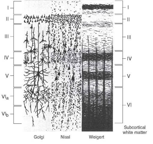
The neocortex have 6 layers and allocortex have only 3 layers.

Cerebral cortex layers microanatomy

The 6 layers of Neocortex:

Orientation of layers:

  • Outer: Towards meaninges
  • Inner: Towards white matter

Idea about the layers:

  • Molecular or plexiform: Only cell processes
  • Granular layer: Densely packed stellate cells
  • Pyramidal layer: Medium and Large pyramidal cells
  • Multiform layer: Different types of cells
  • Pyramidal cells are absent in inner granular layer
Layers Components Afferents Efferents
I – Molecular Axons and Dendrites (Cell processes) From other regions of Cortex and Brainstem To other regions of cortex

(Intra-cortical

Association functions)

II – External granular Densely packed Stellate cells + Small pyramidal cells
III – External pyramidal Loosely packed Stellate cells + Medium pyramidal cells
IV – Internal granular Densely packed Stellate cells only + From Thalamus
V – Internal pyramidal Large pyramidal cells only (few stellate cells) – Giant Pyramidal cells of Betz + From Brain stem To Brain stem & Spinal cord (Projection fibers)
VI – Multiform Multiple sized pyramidal cells + Loosely packed stellate cells To Thalamus

The 3 layers of allocortex:

  1. Molecular layer
  2. Pyramidal layer
  3. Multiform layer

Allocortex is found in: Limbic system

  1. Olfactory cortex
  2. Hippocampal formation
  3. Subiculum

Mesocortex: A transitional type of 3 to 6-layered cortex between neocortex and allocortex –

  1. Parahippocampal gyrus
  2. Pre- and Para-subiculum
  3. Insula

real cortex

Neurons of the cortex:

  1. Pyramidal cells:
    • Large layer V pyramidal cells project axons to brainstem and spinal cord.
    • Smaller layer II and III pyramidal cells project axons to other cortical areas.
  2. Stellate cells: Interneurons whose axons remain within the cortex
  3. Fusiform cells (in deeper layers): Gives rise to corticothalamic projections
  4. Horizontal cells of Cajal
  5. Cells of Martinotti

Bands of Baillarger: Formed by high concentration of horizontally arranged nerve fibers.

  • External band: In layer IV
  • Internal band: In layer V


dr. sulabh kumar shrestha
Dr. Sulabh Kumar Shrestha, MS Orthopedics

He is the section editor of Orthopedics in Epomedicine. He searches for and share simpler ways to make complicated medical topics simple. He also loves writing poetry, listening and playing music. He is currently pursuing Fellowship in Hip, Pelvi-acetabulum and Arthroplasty at B&B Hospital.

14 shares
  • Facebook14
  • Twitter
PGMEE, MRCS, USMLE, MBBS, MD/MS AnatomyNervous system

Post navigation

Previous post
Next post

Related Posts

PGMEE, MRCS, USMLE, MBBS, MD/MS

Kambin’s Triangle or Prism

Apr 10, 2025Apr 10, 2025

There is confusion regarding the borders of Kambin’s triangle across the literature. Kambin originally described the borders of this triangular working zone as: Though it was named as a “triangle” it was described with 4 borders with 2 triangles (in AP view and Lateral view) which gives a 3D structure…

Read More
PGMEE, MRCS, USMLE, MBBS, MD/MS NG insertion technique

NG tube insertion

Dec 1, 2015Jun 18, 2016

Indications Gastric decompression Gavage (feeding) Gastric lavage (irrigation or dilution of gastric contents) Sampling of the gastric contents for analysis Sometimes used to instil air into the stomach to detect a gastric perforation on erect CXR Contraindications Basilar skull fractures (Risk of NG reaching brain) Facial fractures (risk of cribiform…

Read More
PGMEE, MRCS, USMLE, MBBS, MD/MS

Thyroid Gland Blood Supply : Mnemonics

Apr 29, 2024Apr 29, 2024

Arterial supply Arterial anastomosis Venous drainage The venous plexus lies beneath the true capsule (peripheral condensation of fibrous stroma of gland). Hence, during thyroidectomy, thyroid gland is removed along with the true capsule to avert the hemorrhage. False capsule (pre-tracheal fascia) is left intact. Thyroid Arteries and Laryngeal Nerves Mnemonic:…

Read More

Comment

  1. Lilian says:
    Oct 25, 2025 at 12:10 pm

    Clear and Concise- thanks!

    Reply

Leave a Reply Cancel reply

Your email address will not be published. Required fields are marked *

This site uses Akismet to reduce spam. Learn how your comment data is processed.

Shrestha SK. Cerebral Cortex Layers (Microanatomy) Simplified [Internet]. Epomedicine; 2016 Jul 30 [cited 2026 May 20]. Available from: https://epomedicine.com/medical-students/cerebral-cortex-layers-microanatomy-simplified/.

Pre-clinical (Basic Sciences)

Anatomy

Biochemistry

Community medicine (PSM)

Embryology

Microbiology

Pathology

Pharmacology

Physiology

Clinical Sciences

Anesthesia

Dermatology

Emergency medicine

Forensic

Internal medicine

Gynecology & Obstetrics

Oncology

Ophthalmology

Orthopedics

Otorhinolaryngology (ENT)

Pediatrics

Psychiatry

Radiology

Surgery

RSS Ask Epomedicine

  • What to study for Clinical examination in Orthopedics?
  • What is the mechanism of AVNRT?

Epomedicine weekly

  • About Epomedicine
  • Contact Us
  • Author Guidelines
  • Submit Article
  • Editorial Board
  • USMLE
  • MRCS
  • Thesis
©2026 Epomedicine | WordPress Theme by SuperbThemes