INFECTIVE ENDOCARDITIS INDICATIONS FOR SURGERY
- A: Aortic valve, Annular Abscess
- B: Block (heart block), Bacteremia (persistent)
- C: Complications, CCF
- D: Destructive penetrating lesion
- E: Emboli
- F: Fever despite appropriate antibiotics, Fungus
- G: Growing vegetations
- H: Heart failure, Highly resistant organisms
PROSTHETIC HEART VALVES COMPLICATIONS: VIP BEAST
V: Valve Obstruction
I: INR monitoring, Infective Endocarditis
P: Paravalvular regurgitation, Prosthetic mismatch
B: Bleeding
E: Embolization
A: Anemia (the smaller the ring leaks, the severer the hemolysis)
S: Structural failure
T: Thrombosis
CAUSES OF MI WITHOUT CORONARY ATHEROSCLEROSIS – MINOCA: CLEAN VASE
- Clean: Coagulopathies (hypercoagulable states: PRV, Thrombocytosis, Factor V Leiden, PC&S def, APLS)
- V: Vasculitis: SLE, PAN, TA, Kawasaki
- A: Anomalous origin of coronary artery
- S: Spasm: Variant angina, Cocaine, Kounis syndrome (check the 3 types, and do not give beta blockers)
- E: Embolus: Atrial myxoma, A&V thrombus
FORRESTER DIAMOND CLASSIFICATION OF ACUTE HEART FAILURE (because I couldn’t‘t remember what it was where!)
I remember it as the SUN & RAIN classification.
Imagine a house in the top left quadrant. Inside is Dry and Warm. Outside is Wet and Cold
So, on top is warm (because of the sun). At the bottom is cold (because seats outside the house)
To the left is dry (because is inside the house, to the right is wet because it is outside the house and rains (congested)
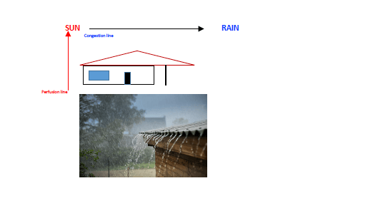
If SBP-DBP < 25%, then CO < 2.2
SOME DIFFERENCES
| S1Q3T3APE | S1q3R3 Emphysema with marked pulmonary HTN | SISIISIII [SII>SIII] The S wave should be greater than the preceding R wave in at least one of these leads RVH basal region |
| S1: Acute Pulmonary Embolism Immediately after the embolic event is broad and shallow, and changes to deep and narrow; usually undergoes rapid resolution Q3 (not pathological): is NOT ³ 0,04 sec or longer in duration & NOT ³ 25% of the ensuing R waveT3: means left axis deviation (-30°) of the T wave axis (simulates acute inferior MI) | In COPD & emphysema there is usually RAD of the frontal plane QRS axis. With the development of marked pulmonary HTN, the QRS axis deviates further to the right clockwise to +120° or even +150°. In very severe cases is directed to the NW region | Anterior Wall Myocardial Infarction (Apical wall – permanent change; rSr’ V1,2= El-Sherif sign) RV dominance [RVH of congenital heart disease & acquired heart disease – cor pulmonale (with RAE)]Straight back syndrome [LAD -120° (»LAFB), rSr’V1, SII>SIIINormal healthy adults |
DIFFERENCE BETWEEN SVT AND V. TACH: THE STUTTER
- A: AV dissociation (confirms V Tach)
- A: Absent BBB
- B: Brugada sign: QS interval > 100 msec
- C: Concordance (+ or -) monophasic
- C: Capture beats
- D: Dilated QRS > 160 msec Josephson sign
- E: Extreme axis deviation: North West axis. Vereckei algorithm, + aVR
- E: Ear rabbit left: Marriot sign
- F: Fusion beats
STEMI MIMICS: when you see VPB (Ventricular Premature Beats) pray a few AVE MARIAS
- V: Voltage
- P: Pacemaker
- B: Brugada
- A: Acute coronary occlusion
- V: Vasospasm [Kounis syndrome & Prinzmetal]
- E: Early repolarization
- M: Myo, pericarditis
- A: Aneurysm, ventricular (Look for El-Sherif sign)
R: Raised K+, Ca2+ - I: Intracranial abnormalities
- A: Acute Pulmonary Embolism
- S: Stress (Tako Tsubo), Spiked Helmet sign
LAW [LEFT ATRIAL WALL] CALCIFICATIONS
- Porcelain LA or Coconut LA
- Porcelain Atrium has implications for MV surgery
- Almost all have atrial fibrillation (average duration 10 years)
- Usually spares inter atrial septum
- 3 types ABC = AFP
- A: Appendage only; almost always associated with LAA thrombus
- B: for Be Free wall of the LA + MV
- C: Confined to Posterior wall. McCollum patch results from a jet lesion 2° to MR
- It may require endoatriectomy @ the time of valve replacement
- Rule out ESKD & Endocarditis
CAUSES OF TR
A: Addict, Attack stabbing
B: BP (Pulm Arterial Hypertension), Blunt trauma
C: CCF, Carcinoid
D: Diseased valve (prolapsed TV)
E: Endocarditis, Ebstein anomaly
F: Fibrosis (endomyocardial fibrosis – very important in East Africa), Following PAH
SCHAMROTH PENTAD for typical HEBERDEN angina: 5 Key questions that Prof Schamroth used to ask to any px presenting w/ chest pain
- One 1 finger vs fist (if px points with one finger, it’s unlikely to be ischemia)
- 2nd wind (pxs with angina after some effort seems to possess a 2nd wind)
- 30” to 30 minutes (lasting; if it lasts > 30’, is not stable angina)
- Work (= 4 letters): usual work does not cause the pain; unusual work lowers the threshold for pain
- Minutes – relief within 5 minutes

Hard worker, Reliable, Team player, Family man.
MBChB (O’Porto Univ.), Dip HIV Man (CMSA), DTM&H (Wits), DipPEC (CMSA), Dip Internal Medicine (CMSA), M. Med. Clinical Pharm [Cum Laude] (Univ. Pretoria), FCP (CMSA)