Skip to content
Epomedicine

Mnemonics, Simplified Concepts & Thoughts

Epomedicine

Mnemonics, Simplified Concepts & Thoughts

renal papillary necrosis patterns

Ball-on-Tee, Lobster Claw and Signet Ring Sign – Renal papillary necrosis

Epomedicine, Oct 27, 2016

Synonym: Necrotizing papillitis

In renal papillary necrosis, part or all of necrotic renal papilla sloughs and may fall into the pelvicalyceal system, which may remain there (and get calcified) or may be voided down the ureter (often causing obstruction). The renal medulla and papillae are vulnerable to ischemic necrosis because of peculiar arrangement of their blood supply and hypertonic environment.

Common causes of papillary necrosis

Remember the mnemonic: POSTCARDS

  • Pyelonephritis
  • Obstruction of the urinary tract
  • Sickle cell hemoglobinopathies, including sickle cell trait
  • Tuberculosis
  • Cirrhosis of the liver, Chronic alcoholism
  • Analgesic abuse
  • Renal transplant rejection, Radiation
  • Diabetes mellitus (commonest – accounts for more than 50% of the cases of renal papillary necrosis 1Radiology Illustrated: Uroradiology edited by Seung Hyup Kim)
  • Systemic vasculitis

Best modality of evaluation of papillary necrosis

IVU or retrograde pyelography

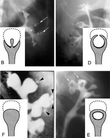
Radiological signs of papillary necrosis

If the papillary sloughing is:

  1. Partial: Contrast tracks around or onto it
  2. Complete: Calyx appears spherical (with loss of papillaryindentation) and as filling defect
necrotizing papillitis signs
Normal (A), Central excavation with ball-on-tee
appearance (B), Forniceal excavation (C), Lobster
claw appearance (D), Signet ring appearance (E),
and sloughed papilla with clubbed calix (F)

Ball-on-tee appearance: Contrast material filling central excavations in the papilla of the interpolar region gives ball-on-tee appearance.

Lobster claw sign: Excavation extending from the caliceal fornices produces the lobster claw deformity.

Signet ring sign: The necrotic papillary tip may remain within the excavated calyx, producing the signet ring sign when the calyx is filled with contrast material.

Club shaped saccular calyx: Due to sloughed papilla

renal papillary necrosis patterns

37 shares
  • Facebook17
  • Twitter
PGMEE, MRCS, USMLE, MBBS, MD/MS Internal medicineRadiologyRenal and Electroloyte

Post navigation

Previous post
Next post

Related Posts

PGMEE, MRCS, USMLE, MBBS, MD/MS

Spinal Tumors Localization : Mnemonics

Apr 9, 2025Apr 9, 2025

Intradural Intramedullary Spinal Tumors Mnemonic: I HEAL Intradural Extramedullary Spinal Tumors Mnemonic: MNM Extradural Spinal Tumors Lesions outside the thecal sac are categorized as extradural lesions. Remember that everything that isn’t in the thecal sac is extradural, including discs, bones, nerves, and blood vessels.

Read More
PGMEE, MRCS, USMLE, MBBS, MD/MS

Osteopetrosis : Mnemonic

Jul 20, 2022Jul 20, 2022

Synonyms: Marble bone disease Albers-Schonberg disease (Autosomal dominant form) Mnemonic: OSTEOPETROSIS Osteoclast dysfunction (Carbonic anhydrase II gene mutation or chloride channel dysfunction) Symmetric skeletal sclerosis Thickened cortex with loss of medullary canal diameter (bone in bone appearance) Extramedullary hematopoiesis (aplastic anemia and hepatosplenomegaly) Overgrowth of skull foramina (cranial nerve palsies)…

Read More
PGMEE, MRCS, USMLE, MBBS, MD/MS wilms tumor

Wilm’s Tumor (Nephroblastoma) : Quick review

Oct 27, 2017Jul 13, 2020

Wilm’s tumor is the most common primary renal tumor of childhood. Origin: Embryonic nephrogenic (mesodermal) cells Mechanism: Deletion of WT-1 gene on chromosome 11 Pathology: a. Macroscopic (Gross): large, solitary tan to grey color mass; pale on cut section with areas of hemorrhage; 10% are bilateral or multicentric; grossly difficult…

Read More

Comment

  1. Manal says:
    Jan 17, 2022 at 6:27 am

    Really very informative and consize

    Reply

Leave a Reply to Manal Cancel reply

Your email address will not be published. Required fields are marked *

This site uses Akismet to reduce spam. Learn how your comment data is processed.

Pre-clinical (Basic Sciences)

Anatomy

Biochemistry

Community medicine (PSM)

Embryology

Microbiology

Pathology

Pharmacology

Physiology

Clinical Sciences

Anesthesia

Dermatology

Emergency medicine

Forensic

Internal medicine

Gynecology & Obstetrics

Oncology

Ophthalmology

Orthopedics

Otorhinolaryngology (ENT)

Pediatrics

Psychiatry

Radiology

Surgery

RSS Ask Epomedicine

  • What to study for Clinical examination in Orthopedics?
  • What is the mechanism of AVNRT?

Epomedicine weekly

  • About Epomedicine
  • Contact Us
  • Author Guidelines
  • Submit Article
  • Editorial Board
  • USMLE
  • MRCS
  • Thesis
©2026 Epomedicine | WordPress Theme by SuperbThemes