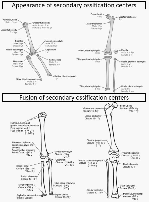
Upper end of humerus
Mnemonic: HGL – 1,3,5
a. Head – by 1 year
b. Greater tuberosity – by 3 years
c. Lesser tuberosity – by 5 years
All 3 centers fuse together by 6 years (1 year after the last center appears)
Upper end of femur
Mnemonic: HGL – 1,4,14
a. Head – by 1 year
b. Greater trochanter – by 4 years
c. Lesser trochanter – by 14 years
Elbow
Mnemonic: CRITOE (Add 2 years in sequence); variation of +/- 1 year
- Capitulum: by 2 years
- Radial head: by 4 years
- Internal (medial) epicondyle: by 6 years
- Trochlea: by 8 years
- Olecranon (Upper end of ulna): by 10 years
- External (lateral) epicondyle: by 12 years
Capitulum, Trochlea and Lateral epicondyle fuses together to form conjoint epiphysis in 14 years and the conjoint epiphysis fuses with the shaft in 15 years.
Medial epicondyle fuses separately with the shaft in 16 years.
Distal radius and Ulna
From the above mnemonic, we know that the time of appearance of proximal radius and ulna is 4-5 years and 9-10 years respectively.
Remember in sequence: 1, 5, 5, 10 (like a letter “N“)
a. Distal radius: by 1 year
b. Proximal radius: by 5 years
c. Distal ulna: by 5 years
d. Proximal ulna: by 10 years
Carpals, Metacarpals and Phalanges of Hand
Carpal bones: Appearance of carpal bones ossification center : Mnemonic
Secondary ossification centers of Metacarpals and Phalanges appear by 3 years and fuses with the shaft by 18-20 years.
Knee
Distal femur: By birth
Proximal tibia: At birth
Patella: by 4 years (attains adult contour at 14-16 years)
Proximal fibula: by 4 years
Distial tibia and fibula
Both appear by 1 year.
Medial malleolus forms from distal tibial epiphysis at 7-10 years.
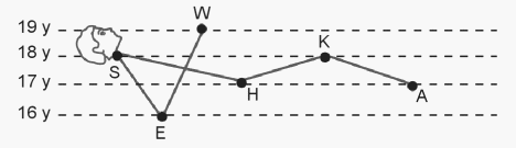
Mnemonic for fusion or Union of epiphyses with shaft in long bones: Imagine a person reclining on a sandy beach as shown in the diagram above. His elbows are sinking in the sand somewhat. Now draw four lines parallel to the horizontal plane. The lowest one passes through the elbow, the next through the hip and ankle joints, the next through the shoulder and knee joint, and the uppermost through the wrist joint. Epiphyses falling on a particular line fuse at a particular age. Thus all epiphyses around the elbow joint complete fusion by 16 years, all epiphyses around hip and ankle joints by 17 years, all epiphyses around shoulder and knee by 18 years, and all epiphyses around the wrist by 19 years.
Tarsals, Metatarsal and Phalanges of foot
Primary ossification centers of cuboid and cuneiform:
Mnemonic: 0, 1, 3, 2 from lateral to medial
- Cuboid: by birth
- Lateral cuneiform: by 1 year
- Intermediate cuneiform: by 3 years
- Lateral cuneiform: by 2 years
Secondary ossification centers of navicular, calcaneus, metatarsal and phalanges:
- Navicular, metatarsal, phalanges: 3 years
- Calcaneus: 5 years
Fusion of metatarsal and phalangeal epiphysis with shaft occurs in 17-18 years.
Sternum
Centers for manubrium sterni and 4 sternebrae (body of sternum) appear sequentially from top to bottom and are present by birth.
Xiphisternum appear by 3 years.
4 sternebrae unite sequentially from top to bottom in 15, 20 and 25 years respectively.
Xiphisternum unites with body of sternum at 40 years.
Manubrium sterni unites with body of sternum at 60-70 years.
Scapula
a. Center of coracoid: 1 year
b. Base of coracoid: 10 years
These 2 centers unite with the scapula at 15 years.
Tip of coracoid: Variable; often at puberty (may fail to fuse)
2 centers of acromion, medial border and inferior angle: appear at puberty and fuse at around 22 years
Hip bone
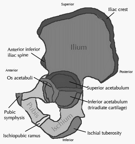
Ischiopubic ramus unites by 7-8 years
Iliac crest and tip of pubis: appear at 14 years and unite by 20 years (union in iliac crest occurs from before backwards)
Tip of ischium: appear at 16 years and unite by 20 years
Tri-radiate cartilage: Ossification in the acetabular cup begins from two separate centers (os acetabuli) between the ilium and pubis, and between the ilium and ischium. As bone begins to be laid from these two centers, the acetabular cup assumes the shape of a triradiate cartilage, which becomes noticeable in radiographs by 13 years and disappears by 15 years.
Sacrum
Body of sacral vertebra begin to unite marginally: by 20 years
Sacrum is one bone: by 25 years (center may remain unossified upto 50 years)
Sources of information:
1. APC Essentials of Forensic Medicine and Toxicology By Anil Aggrawal
2. Radiopedia
Hey can you tell me from which book has that ‘man lying in sand’ photo been taken from?