Skip to content
Epomedicine

Mnemonics, Simplified Concepts & Thoughts

Epomedicine

Mnemonics, Simplified Concepts & Thoughts

Refracture of the Patella – Early TBW Failure and Re-fixation

Dr. Sulabh Kumar Shrestha, MS Orthopedics, Mar 6, 2026Mar 6, 2026

One month following an index TBW procedure (using 4 K‑wires + cerclage), our patient sustained a refracture of the patella after a fall. There was no high-energy direct trauma. This case typifies early failure of a classic TBW construct.

Failure Factors Identified in Literature

  • Selçuk et al. (2025) found a 28% failure rate in TBW, with increased risk linked to single-knot cerclage, long K-wires (>69 mm), and lateral K‑wire placement >10.85 mm from articular surface
  • Surgeons should aim for a K-wire length not exceeding 69 mm and a lateral K-wire to articular distance below 10.85 mm to minimize mechanical insufficiency.

Revision Fixation

  • Employed tension-band wiring with cerclage augmentation, providing redundancy and rigidity
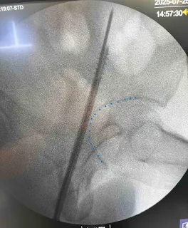
  • The horizontal laceration overlapped with the previous incision; we extended it to reach the fracture site, preserving soft tissue integrity.

Alternate Techniques

double TBW
Double TBW
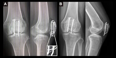
  • Double tension-band wiring with cerclage augmentation consistent with Xue et al.’s revision technique that yields 100% union rates and excellent function
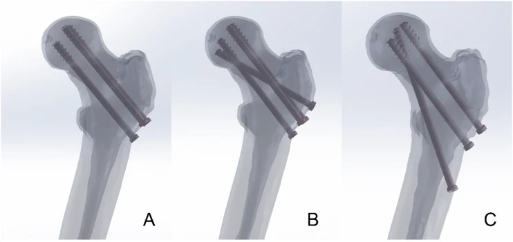
    • Insert 2 2.5-mm Kirschner (K)-wires distally from the proximal pole of the patella through the fracture site and the cortex of the distal fragment
    • A stab incision is made at each end of the K-wires to expose the surface of the bone
    • Two stainless steel wires is looped around the K-wires separately in a figure-eight manner.
    • Tighten the loops to make them cling to the anterior surface of the patella and twist the steel wires at the insertions in the K-wires
    • Cerclage wiring is then performed using a cable or a stainless steel wire
  • For severely comminuted or lower‑pole fractures, non-absorbable braided suture with circumferential wiring can be an effective alternative achieving union in 14 weeks and minimizing metal hardware issues

Key Takeaways for Surgeons

  • Early refracture (<6 weeks) often signals poor initial construct stability
  • Critical technical factors include knot configuration, appropriate K‑wire length, and optimal wire placement relative to patellar anatomy
  • A dual-band TBW with cerclage augmentation offers stronger fixation, especially in revision scenarios
  • Alternate suture-based fixation can be useful in comminuted or soft-tissue compromised fractures
dr. sulabh kumar shrestha
Dr. Sulabh Kumar Shrestha, MS Orthopedics

He is the section editor of Orthopedics in Epomedicine. He searches for and share simpler ways to make complicated medical topics simple. He also loves writing poetry, listening and playing music. He is currently pursuing Fellowship in Hip, Pelvi-acetabulum and Arthroplasty at B&B Hospital.

  • Facebook
  • Twitter
Fellowship Blog OrthopedicsTrauma

Post navigation

Previous post
Next post

Related Posts

Fellowship Blog

Mathematical Principles in Mechanically Aligned Total Knee Arthroplasty

Mar 21, 2026Mar 21, 2026

Mechanical Axis Concept Natural Knee Target alignment in MA-TKA Tibial cut Distal femoral cut Posterior femoral cut The 3° Compensation Rule Distal femoral and Tibial resection thickness = Implant thickness 2 mm rule in TKA References: Dr. Sulabh Kumar Shrestha, MS Orthopedics He is the section editor of Orthopedics in…

Read More
Fellowship Blog

Normal Hip Development

Dec 23, 2025Feb 2, 2026

4-6 weeks of gestation: By 7 weeks of gestation: By 11 weeks of gestation: Late gestation: Postnatal: The acetabulum is deepened by the natural pressure from the developing femoral head on the acetabulum. Hips in newborns with developmental dysplasia are not just normal hips with capsular laxity; they are structurally…

Read More
Fellowship Blog

The 0-3-6-9 rule in Mechanical Alignment Total Knee Arthroplasty (MA-TKA)

Feb 25, 2026Feb 25, 2026

The goal of MA-TKA is to restore the normal mechanical axis. In contrast to kinematic axis alignment, which attempts to recreate native knee joint line (3° tibial varus and 3° femoral valgus), in MA-TKA, the distal femur and the tibia are cut to be perpendicular (0°) to the mechanical axis….

Read More

Leave a Reply Cancel reply

Your email address will not be published. Required fields are marked *

This site uses Akismet to reduce spam. Learn how your comment data is processed.

Pre-clinical (Basic Sciences)

Anatomy

Biochemistry

Community medicine (PSM)

Embryology

Microbiology

Pathology

Pharmacology

Physiology

Clinical Sciences

Anesthesia

Dermatology

Emergency medicine

Forensic

Internal medicine

Gynecology & Obstetrics

Oncology

Ophthalmology

Orthopedics

Otorhinolaryngology (ENT)

Pediatrics

Psychiatry

Radiology

Surgery

RSS Ask Epomedicine

  • What to study for Clinical examination in Orthopedics?
  • What is the mechanism of AVNRT?

Epomedicine weekly

  • About Epomedicine
  • Contact Us
  • Author Guidelines
  • Submit Article
  • Editorial Board
  • USMLE
  • MRCS
  • Thesis
©2026 Epomedicine | WordPress Theme by SuperbThemes