Skip to content
Epomedicine

Mnemonics, Simplified Concepts & Thoughts

Epomedicine

Mnemonics, Simplified Concepts & Thoughts

pulmonary embolism xray

Pulmonary Embolism : Chest X-ray Signs

Epomedicine, Oct 25, 2017

pulmonary embolism xray

1. Classic presentation is normal X-ray in patient with dyspnea and hypoxia

2. Atelectasis or parenchymal abnormality (68%)

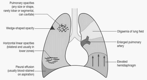
3. Elevated hemidiaphragm

4. Pleural effusion (Felson’s sign – pleural effusion on left > right)

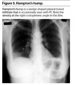
5. Hampton’s hump: peripheral pleural based wedge-shaped density above the diaphragm due to pulmonary infarct

hamptons hump

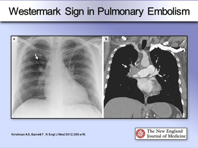
6. Westermark’s sign: is distension of pulmonary vasculature proximal to embolism with loss of vascular markings distally, i.e. localized peripheral oligemia (rare)

westermark sign

7. Palla sign: Enlarged right descending pulmonary artery

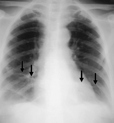
8. Fleischner lines: Long curvilinear densities reaching pleural surface

fleischner lines

9. Fleischner sign: Dilated pulmonary artery

84 shares
  • Facebook82
  • Twitter
Emergency Medicine PGMEE, MRCS, USMLE, MBBS, MD/MS Emergency medicineInternal medicinePediatricsRadiologyRespiratory system

Post navigation

Previous post
Next post

Related Posts

PGMEE, MRCS, USMLE, MBBS, MD/MS

Superior Orbital Fissure (SOF): Mnemonics

Aug 5, 2023Aug 5, 2023

Superior Orbital Fissure (SOF) is a cleft between greater and lesser wing of sphenoid which connects orbit and middle cranial fossa. It is divided into 3 compartments by the tendinous ring (annulus of Zinn). Superior orbital fissure alone (Lateral compartment): Mnemonic: LFTS Superior orbital fissure in tendinous ring (Intermediate compartment):…

Read More
PGMEE, MRCS, USMLE, MBBS, MD/MS schatzker classification

Schatzker classification for Tibial plateau fracture : Mnemonic

Apr 15, 2020Aug 18, 2021

Schatzker system is widely accepted and used to classify tibial plateau fractures. It is based on a 2D representation of the fracture. As a general rule, it can be divided into low-energy and lateral tibial plateau variants (types I-III) and high energy variants (type IV-VI). However, it does not include…

Read More
PGMEE, MRCS, USMLE, MBBS, MD/MS

Differentiating Necrotizing Fasciitis from other soft tissue infections

Sep 30, 2020Oct 2, 2020

Necrotizing fasciitis can be misdiagnosed in about 75% of the cases in the intial stage of the disease. The most consistent feature of early necrotizing fasciitis is the pain out of proportion to swelling or erythema. Other features helping to differentiate from other soft tissue infections are: Tenderness extending beyond…

Read More

Comment

  1. Dr Gebeyehu Bekele says:
    Apr 3, 2023 at 5:20 pm

    It is very important summary,thanks

    Reply

Leave a Reply Cancel reply

Your email address will not be published. Required fields are marked *

This site uses Akismet to reduce spam. Learn how your comment data is processed.

Epomedicine. Pulmonary Embolism : Chest X-ray Signs [Internet]. Epomedicine; 2017 Oct 25 [cited 2025 Dec 6]. Available from: https://epomedicine.com/emergency-medicine/pulmonary-embolism-chest-x-ray-signs/.

Pre-clinical (Basic Sciences)

Anatomy

Biochemistry

Community medicine (PSM)

Embryology

Microbiology

Pathology

Pharmacology

Physiology

Clinical Sciences

Anesthesia

Dermatology

Emergency medicine

Forensic

Internal medicine

Gynecology & Obstetrics

Oncology

Ophthalmology

Orthopedics

Otorhinolaryngology (ENT)

Pediatrics

Psychiatry

Radiology

Surgery

RSS Ask Epomedicine

  • What to study for Clinical examination in Orthopedics?
  • What is the mechanism of AVNRT?

Epomedicine weekly

  • About Epomedicine
  • Contact Us
  • Author Guidelines
  • Submit Article
  • Editorial Board
  • USMLE
  • MRCS
  • Thesis
©2025 Epomedicine . All rights reserved.