Skip to content
Epomedicine

Mnemonics, Simplified Concepts & Thoughts

Epomedicine

Mnemonics, Simplified Concepts & Thoughts

Trendelenburg sign test

Trendelenburg test or sign

Epomedicine, Jun 4, 2014Sep 5, 2022

Method: The patient is then asked to stand on one leg and bend the opposite knee to 90° without flexing the hip. This action eliminates the role of hip flexors as they play a role in pelvic stability and may affect the Trendelenburg sign. The test is then repeated by standing on the other leg. The sign is deemed negative, i.e. the abductor mechanism is normal, when the pelvis remains level. A positive result indicates abductor mechanism dysfunction and the pelvis on the unsupported side will descend. A patient standing on his right leg would be Trendelenburg-positive for the right if the left side dipped.

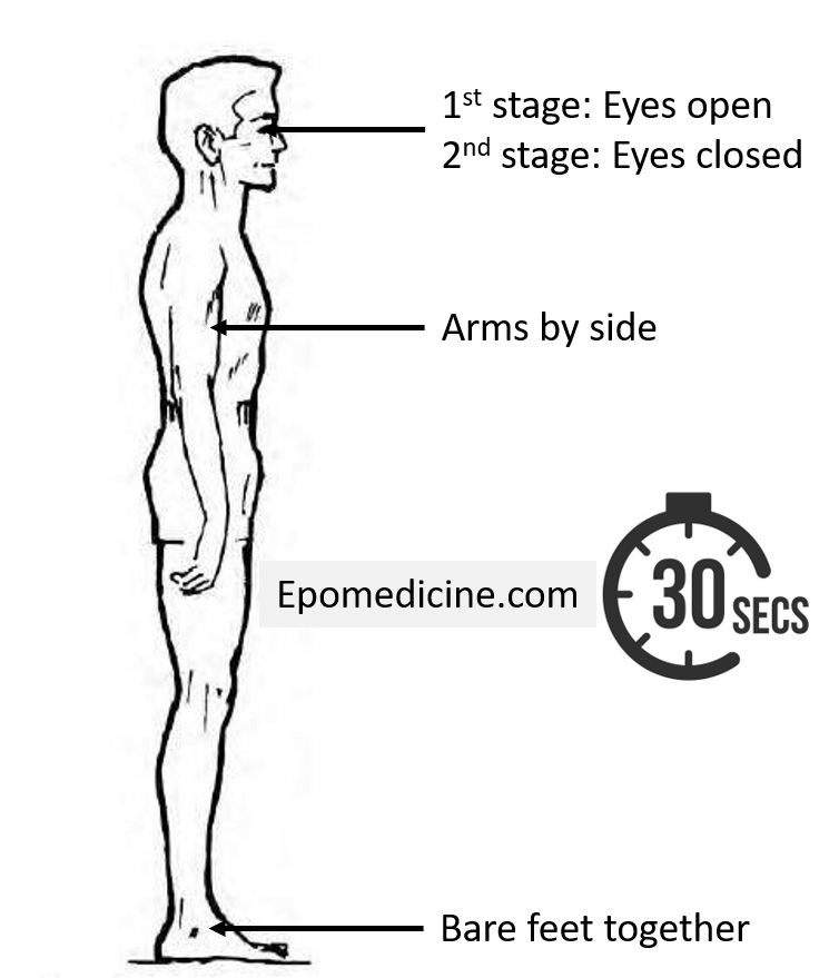
Delayed Trendelenburg test: Similar to the Trendelenburg test but the patient is made to stand for 30 seconds to assess the abductor’s fatigue as in acetabular dysplasia. However, it’s value has been questioned as any painful hip condition may produce positive results.

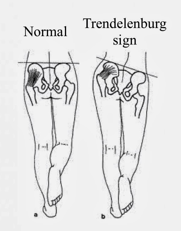
Normal: When the body weight is supported on one limb, the abductors (gluteus medius and minimus) of the supported side contract and raise the opposite unsupported side of the pelvis.

Abnormal: If the abductor mechanism is defective, the unsupported side of pelvis drops and this is known as positive trendelenburg’s test. Patient may not descend on one side when assessing for the Trendelenburg sign, as they can compensate by leaning over the affected hip to shift the body’s center of gravity in that direction (Trendelenburg lurch).

Mnemonic: Sound Side Sags

Use: To assess the integrity of abductor mechanism of hip

Causes of positive trendelenburg sign or test:

This test is positive in conditions in which any of the three – fulcrum (femoral head), lever arm (femoral neck length) or power (muscles/nerves) are affected.

Note: The primary abductors of hip are gluteus medius and minimus while tensor fascia lata and sartorius are accessory abductors of hip.

Trendelenburg sign test

1. Power paralysis of abductor muscles:

  • Superior gluteal nerve palsy
  • Polio
  • Iliotibial tract palsy

2. Decreased lever arm:

  • Fracture neck of femur

3. Absence of stable fulcrum:

  • Dislocation of hip
  • Destruction of femoral head: Perthe’s disease, Avascular necrosis of head of femur, Late stages of TB hip, Septic arthritis

Pathomechanics of Trendelenburg sign:

When people walk, their pelvises don’t drop dramatically on the non-stance side because gravity’s adductor moment is balanced by an equal and opposite abductor moment. In single leg stance, the effective center of gravity shifts to the non-supportive leg and a downward force attempts to tilt pelvis on unsupported side. The abductors on the hip which supports the body, must exert a downward counter balancing force with right hip joint acting as a fulcrum. Weakness of the abductor leads to sagging of pelvis on unsupported side.

41 shares
  • Facebook
  • Twitter
Clinical Skills and Approaches Clinical examinationOrthopedics

Post navigation

Previous post
Next post

Related Posts

Clinical Skills and Approaches

Donning and Doffing Sequence – Mnemonic

Jun 7, 2020Jun 7, 2020

It’s Covid-19 pandemic and it seems like Covid is here to stay for a while. Donning (putting on) and Doffing (taking off) of PPE (Personal Protective Equipment) have suddenly gained a great importance for the health professionals and many of us must have already practised this. RN Susan E. Sammons…

Read More
Clinical Skills and Approaches relative bradycardia enteric fever

Relative Bradycardia

Nov 1, 2017

Synonyms: Faget sign, Sphygmo-thermic dissociation, Sphygmo-thermal dissociation Definition of Relative Bradycardia Physiologically, for each 1 °F rise in body temperature, there is a commensurate increase in the heart rate of 10 beats/min . When temperature elevations are not accompanied by a physiologic increase in the pulse, the patient is said…

Read More
Clinical Skills and Approaches spleen percussion

Percussion of Spleen

Jan 31, 2017

Traube’s semilunar space Borders: Superiorly: Left 6th rib superiorly Laterally: Left midaxillary line or Left anterior axillary line Inferiorly: Left costal margin Method: Patient’s position: supine with left arm slightly abducted. Percuss: from medial to lateral Interpretation: Resonance (Normal) and Dullness (Splenomegaly) Also: Pleural effusion or mass in stomach may…

Read More

Comment

  1. palash says:
    May 31, 2015 at 3:12 am

    Text is consized and helpful. Video is very ordinary and not making any sense,

    Reply

Leave a Reply Cancel reply

Your email address will not be published. Required fields are marked *

This site uses Akismet to reduce spam. Learn how your comment data is processed.

Epomedicine. Trendelenburg test or sign [Internet]. Epomedicine; 2014 Jun 4 [cited 2025 Dec 23]. Available from: https://epomedicine.com/clinical-medicine/trendelenburg-test-or-sign/.

Pre-clinical (Basic Sciences)

Anatomy

Biochemistry

Community medicine (PSM)

Embryology

Microbiology

Pathology

Pharmacology

Physiology

Clinical Sciences

Anesthesia

Dermatology

Emergency medicine

Forensic

Internal medicine

Gynecology & Obstetrics

Oncology

Ophthalmology

Orthopedics

Otorhinolaryngology (ENT)

Pediatrics

Psychiatry

Radiology

Surgery

RSS Ask Epomedicine

  • What to study for Clinical examination in Orthopedics?
  • What is the mechanism of AVNRT?

Epomedicine weekly

  • About Epomedicine
  • Contact Us
  • Author Guidelines
  • Submit Article
  • Editorial Board
  • USMLE
  • MRCS
  • Thesis
©2025 Epomedicine . All rights reserved.