Skip to content
Epomedicine

Mnemonics, Simplified Concepts & Thoughts

Epomedicine

Mnemonics, Simplified Concepts & Thoughts

Autonomous zones

Autonomous Sensory Zones of Peripheral Nerves

Epomedicine, May 12, 2014May 12, 2014

Synonym: Autonomous area

Definition: These are the regions where single nerve roots supply distinct and non-overlapping areas of skin. By their nature the “autonomous zones” represent only a small portion of any dermatome and only a few nerve roots have such autonomous zones. The size of autonomous zone for a particular nerve is variable from individual to individual.

Sensory zones of a Peripheral nerve:

  1. Maximal zone: Maximal area supplied by a peripheral nerve
  2. Intermediate zone: Area of overlap of the maximal zone of different peripheral nerves
  3. Autonomous zone: Area exclusively supplied by a particular peripheral nerve

Note:

  • Maximal zone = Intermediate zone + Autonomous zone
  • With the interruption of a sensory nerve, all modalities of cutaneous sensation are lost only over the autonomous zone.
  • Within a few days of complete interruption of a sensory nerve, the autonomous zone shrinks and the intermediate zone enlarges, which may be due to actual ingrowth of fibers from adjacent normal nerve.

Autonomous zones of various nerves:
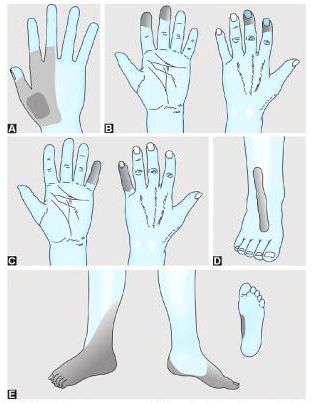
Autonomous zones
A. Radial nerve
B. Median nerve
C. Ulnar nerve
D. Common peroneal nerve
E. Sciatic nerve

  1. Radial nerve: 1st dorsal web space of hand (Anatomical snuff box)
  2. Median nerve: Distal phalanx (tip) of index finger (2nd finger)
    • Other: Tip of thumb
  3. Ulnar nerve: Distal phalanx (tip) of little finger (5th finger)
  4. Common peroneal nerve/Common fibular nerve/External popliteal nerve/Lateral popliteal nerve: Central strip on dorsum of foot
  5. Deep peroneal nerve: 1st dorsal web space
  6. Posterior tibial nerve: Sole of foot
  7. Sciatic nerve: Mixed pattern of Common peroneal nerve and Posterior tibial nerve

Note: According to some authors, radial nerve and common peroneal do not have autonomous zones although complete transection of the nerve results in sensory loss over the above mentioned regions.

MRC grading of sensory recovery tested on autonomous zone:

  • S0: Absense of sensibility in the area
  • S1: Recovery of deep cutaneous pain
  • S2: Partial recovery of protective sensation (superficial cutaneous pain and touch)
  • S3: Recovery of protective sensation with accurate localiztion; sensitivity (and hypersensitivity) to cold is usual
  • S4: Recovery of 2 point discrimination (In hand < 8 mm)
  • S5: Normal sensation or Complete recovery
2 shares
  • Facebook2
  • Twitter
Clinical Skills and Approaches Clinical examinationGeneral SurgeryNervous systemOrthopedics

Post navigation

Previous post
Next post

Related Posts

Clinical Skills and Approaches

Communication Module for Obtaining Informed Consent

May 26, 2024May 26, 2024

The basic structure for communication skills remains the same. Recall the mnemonic P I CX3. Take into considerations of following points during consultation. Mnemonic: CONSENTS Mnemonic Headings Comments C Condition & natural history Explain the clinical condition & prognosis O Options & alternatives No treatment, conservative, medical, radiological, surgicalMake patients aware…

Read More
Clinical Skills and Approaches

Management of Diabetes – GLUCOSE BAD mnemonic

Jul 25, 2020Jul 25, 2020

Glycemic control HbA1c 3 monthly or Bianually (if treatment goal acheived and glycemic control considered stable) – Target: Non-pregnant adult patients: <7% Minor cardiovascular disease, minor hypoglycemic episodes, short diagnosis time with diabetes, and long life expectancy: <6.5% Significant co-morbidities, progressive micro- and macrovascular complications, or short life expectancy: <8%…

Read More
Clinical Skills and Approaches history and examination format

Cyanosis

Dec 3, 2013Feb 16, 2018

Synonyms: Morbus coeruleus, Blue disease Definition of Cyanosis Cyanosis is the bluish discoloration of the skin, mucous membrane and nail bed usually owing to atleast 5 gm/dl of reduced hemoglobin/deoxyhemoglobin or abnormal hemoglobin derivatives (eg. methemoglobin, sulfhemoglobin, etc.) circulating within the superficial dermal capillaries and subpapillary venous plexus (not, as commonly taught, the…

Read More

Leave a Reply Cancel reply

Your email address will not be published. Required fields are marked *

This site uses Akismet to reduce spam. Learn how your comment data is processed.

Pre-clinical (Basic Sciences)

Anatomy

Biochemistry

Community medicine (PSM)

Embryology

Microbiology

Pathology

Pharmacology

Physiology

Clinical Sciences

Anesthesia

Dermatology

Emergency medicine

Forensic

Internal medicine

Gynecology & Obstetrics

Oncology

Ophthalmology

Orthopedics

Otorhinolaryngology (ENT)

Pediatrics

Psychiatry

Radiology

Surgery

RSS Ask Epomedicine

  • What to study for Clinical examination in Orthopedics?
  • What is the mechanism of AVNRT?

Epomedicine weekly

  • About Epomedicine
  • Contact Us
  • Author Guidelines
  • Submit Article
  • Editorial Board
  • USMLE
  • MRCS
  • Thesis
©2026 Epomedicine | WordPress Theme by SuperbThemes