Skip to content
Epomedicine

Mnemonics, Simplified Concepts & Thoughts

Epomedicine

Mnemonics, Simplified Concepts & Thoughts

Surgical landmarks for identification of facial nerve in Parotid surgery

Epomedicine, Dec 31, 2013

The facial nerve (CN VII) exits the skull base through the stylomastoid foramen situated posterolaterally to the styloid process and anteromedially to the mastoid process and enters the parotid gland to divide it into a large superficial lobe and a small deep lobe. The main trunk of facial nerve enters the parotid gland and divides into zygomaticotemporal and cervicofacial divisions at pes anserinus situated approximately 1.3 cm from stylomastoid foramen. Facial nerve paralysis is a daunting potential complication of parotid surgery and is widely reported. Knowledge of the key landmarks of the facial nerve trunk is essential for safe and effective surgical intervention in the region of the parotid gland.

Surgical landmarks to identify main trunk of facial nerve:

1. Tragal “pointer” (of Conley): facial nerve lies approximately 1 cm deep and slightly anterior and inferior to this “pointer”.

  • As the tragal cartilage is dissected free from the parotid fascia, it takses bluntlu pointed shape in its medial aspect, which is called the “pointer”.
  • It is regarded as the most important landmark.
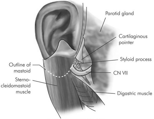
facial nerve parotidectomy

2. Tendon of posterior belly of digastric muscle: facial nerve lies approximately 1 cm deep to the media attachment of the posterior belly of digastric muscle to the digastric groove of mastoid bone.

  • During surgery, the tail of parotid gland is separated from the sternocleidomastoid muscle and the deep underlying posterior belly of digastric muscle, and then the gland is elevated and rotated off of the muscle.

3. Tympanomastoid suture/fissure: facial nerve lies 6-8 mm deep to this suture

  • The tympanomastoid suture is situated at the apex of vagino-mastoid angle or valley of nerve where the mastoid and the vaginal portion of tympanic ring of external ear canal (temporal bone) meets.

4.  Styloid process: lateral to styloid process

Note: 

a. If the proximal segment of facial nerve is obscured, retrograde dissection of 1 or more of the peripheral branches may be necessary to identify the main trunk.

  1. Ramus frontalis is located by a line from tragus to lateral canthus.
  2. Ramus buccalis is located by a line from the tragus towards alae of the nose parallel to the zygoma but 1 cm below.
  3. Ramus mandibularis is near the angle of mandible at a point 4-4.5 cm from the attachment of the lobule of pinna.

b. When necessary, the facial nerve can be identified in the mastoid bone by mastoidectomy and followed peripherally.
75 shares
  • Facebook74
  • Twitter
PGMEE, MRCS, USMLE, MBBS, MD/MS General SurgeryNervous systemOtorhinolaryngology

Post navigation

Previous post
Next post

Related Posts

PGMEE, MRCS, USMLE, MBBS, MD/MS

BIND score in severe hyperbilirubinemia

Oct 27, 2022Oct 27, 2022

Bilirubin-induced neurologic dysfunction (BIND) Score is used to assess bilirubin induced encephalopathy in neonates with severe hyperbilirubinemia. Johnson et al developed the BIND score to help identify an infant who requires more aggressive monitoring and management. The scoring system has 3 parameters: 1. Cry pattern 2. Behavior and mental status…

Read More
PGMEE, MRCS, USMLE, MBBS, MD/MS ulnar nerve course

Ulnar nerve Anatomy – Course and Innervation

Jun 7, 2018Jun 1, 2024

Origin: C(7), C8, T1 (medial cord of Brachial plexus) Course: Motor innervation: 1. Forearm: Flexor carpi ulnaris (weakness of ulnar deviation and flexion of wrist), Medial half of flexor digitorum profundus (branches near the elbow) 2. Hand: branches near wrist Sensory innervation: Palmar branch at forearm and Digital branch at wrist…

Read More
PGMEE, MRCS, USMLE, MBBS, MD/MS

Blood supply of femoral head

May 29, 2020Jan 11, 2024

Three sources of Blood supply Vascular arragement of Proximal Femur a. Extracapsular ring (At the base of femoral neck) These circumflex femoral arteries branch from: Trochanteric anastomosis (centered on trochanteric fossa): Descending branch of SGA + ascending branch of LFCA, MFCA and IGA Cruciate anastomosis (centered on lesser trochanter): Descending…

Read More

Leave a Reply Cancel reply

Your email address will not be published. Required fields are marked *

This site uses Akismet to reduce spam. Learn how your comment data is processed.

Epomedicine. Surgical landmarks for identification of facial nerve in Parotid surgery [Internet]. Epomedicine; 2013 Dec 31 [cited 2026 Aug 24]. Available from: https://epomedicine.com/medical-students/surgical-landmarks-for-identification-of-facial-nerve-in-parotid-surgery/.

Pre-clinical (Basic Sciences)

Anatomy

Biochemistry

Community medicine (PSM)

Embryology

Microbiology

Pathology

Pharmacology

Physiology

Clinical Sciences

Anesthesia

Dermatology

Emergency medicine

Forensic

Internal medicine

Gynecology & Obstetrics

Oncology

Ophthalmology

Orthopedics

Otorhinolaryngology (ENT)

Pediatrics

Psychiatry

Radiology

Surgery

RSS feed: Ask Epomedicine Ask Epomedicine

  • What to study for Clinical examination in Orthopedics?
  • What is the mechanism of AVNRT?

Epomedicine weekly

  • About Epomedicine
  • Contact Us
  • Author Guidelines
  • Submit Article
  • Editorial Board
  • USMLE
  • MRCS
  • Thesis
©2026 Epomedicine | WordPress Theme by SuperbThemes