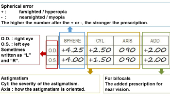
A phoropter/refractor is an instrument commonly used by eye care professionals during an eye examination, containing different lenses used for refraction of the eye during sight testing, to measure an individual’s refractive error and determine his or her eyeglass prescription. Retinoscopy is done instead in children who are unable to…
Tag: Ophthalmology
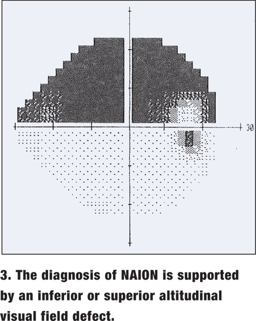
Sudden Vision Loss : Simplified Approach
Acute or sudden vision loss is due to one of the following causes: Opacification of normally transparent structures anterior to retina Retinal abnormalities Abnormalities of optic nerve and visual pathway Systematic history and ocular examination is necessary. Step 1: Unilateral or Bilateral Sudden vision loss ? Monocular loss of vision:…
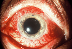
Acute Red Eye : Simplified Approach
Red eye reflects hyperemia or engorgement of superficial visible conjunctival, episcleral or ciliary vessels. A) DIFFERENTIAL DIAGNOSES FOR ACUTE RED EYE 1. Painless red eye: a) Diffuse redness: Lids normal: Conjunctivitis Lids abnormal: Blepharitis Ectropion Trichiasis Eyelid lesion b) Localized redness: Pterygium Corneal foreign body Ocular trauma Subconjunctival hemorrhage Episcleritis
History and Examination in Ophthalmology
A) HISTORY Besides chief complaints, other portion of history is similar to the one you prepare in internal medicine. 1. Description of symptom (SOCRATES): S – Site (Unilateral or Bilateral) O- Onset C – Character R – Radiation (if applicable) A – Aggravating and relieving factors T – Timing and…
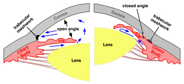
Glaucoma basics : Classification of Glacuoma
Definition: Glaucoma is a group of disorders characterized by a progressive optic neuropathy resulting in a characterstic appearance of the optic disc and a specific pattern of irreversible visual field defects that are associated frequently but not invariably with raised intraocular pressure (IOP). It is one of the commonest cause of irreversible blindness in the world. Open-angle…
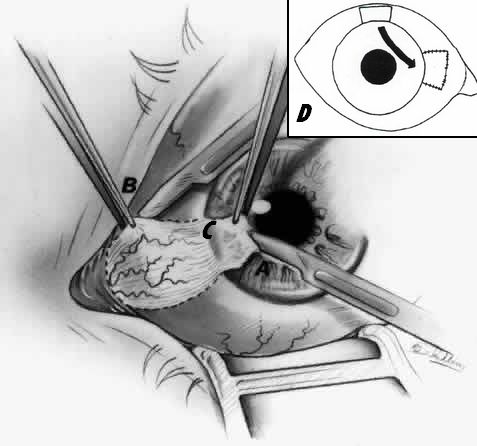
Pterygium Excision
Indications for surgery: 1. Reduced vision secondary to: Threatens visual axis: Pterygium advancing toward or already impinging visual axis Induced astigmatism: Due to flattening of meridian of pterygium by fibrosis in atrophic stage 2. Cosmesis 3. Significant discomfort not relieved by medical therapy 4. Diplopia: Resulting from limited ocular motility…
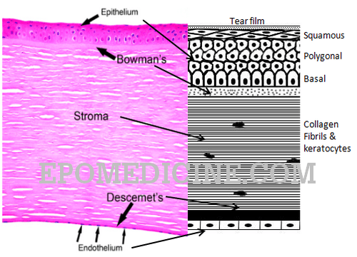
Histology of Cornea and Corneal Dystrophies
Prefix: kerat- Definition: The cornea is a transparent, avascular, watch-glass like structure which forms anterior one-sixth of the outer fibrous coat of the eyeball and covers iris, pupil and anterior chamber. Histology: It consists of 5 distinct layers which can be remembered using the mnemonic “ABCDE“:
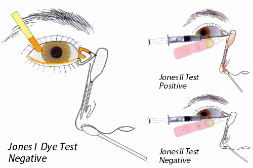
Uses of Fluorescein strips
Content: 1-2% fluorescein dye impregnated Method of use: Explain the procedure to the patient Ask the patient to look up Moisten the strip with a small amount of normal saline or anesthetic drops, without touching the dye impregnated end of the strip with dropper Gently touch the inside of the…
