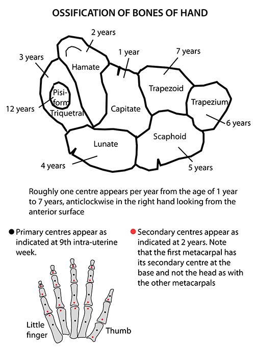
Roughly one center appears per year from the age of 1 year to 7 years, anti-clockwise in right hand and clock-wise in left hand looking from the anterior surface, i.e. from ulnar side to radial side. Pisiform, being a sesamoid bone it gets left behind and only develops years later. capitate: 1-3 months hamate:…
Tag: Musculoskeletal system
Clinical Skills and Approaches
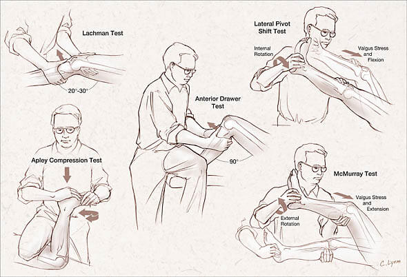
Tests for Knee Ligaments
Anterior Cruciate Ligament (ACL) Lacchman’s test It is performed with the patient supine and the knee flexed 20–30°. The examiner grasps the distal femur (from lateral side) with one hand and the proximal tibia with the other hand (from medial side). The lower leg is given a brisk forward tug…
Emergency Medicine
Ligament Tests for Ankle Injuries
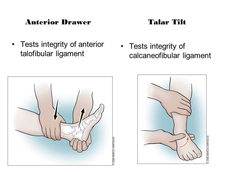
Anterior Drawer Test Assesses: Anterior talofibular ligament (ATFL) Position: Knee joint in flexion and ankle in 10-15 degrees plantar flexion Maneuver: The examiner exerts a downward force on the tibia while simultaneously attempting to “lift up” the foot while grasping behind the heel. Interpretation: A significant difference from the unaffected…
PGMEE, MRCS, USMLE, MBBS, MD/MS
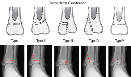
Salter Harris Classification for Physeal Fracture: Mnemonic
In children, the growth plate (physis) is a zone of cartilate situated between the epiphysis and the metaphysis of long bones. Cartilage is weaker than bone and thus is a common site of fracture. The Salter-Harris classification consists of 5 different types of growth plate fractures based on the location…
Emergency Medicine

Ottawa Foot, Ankle and Knee rules – Mnemonic
Ottawa Ankle and Foot Rules Mnemonic: 44-55-66-PM Patients need an X-ray only if: 4: Unable to do 4 steps immediately AND4: Unable to do 4 steps at Emergency Department OR 5: Has pain at the base of 5th metatarsal5: Has pain at the 5caphoid (Navicular) OR 6: Tenderness in 6…
PGMEE, MRCS, USMLE, MBBS, MD/MS
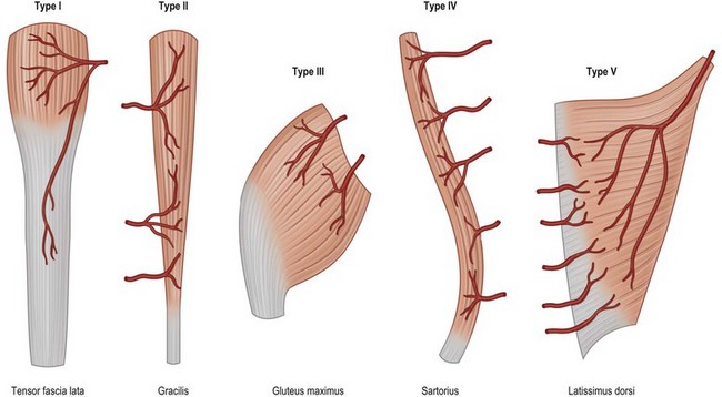
Mathes and Nahai Classification of Muscle Flap based on Vascular Anatomy
Type Dominant pedicle Minor pedicle Example I 1 – Tensor Fascia Lata (TFL) II 1 1 Gracilis III 2 – Gluteus maximus IV – Multiple Sartorius V 1 Multiple Lattisimus dorsi Mnemonic: Ten Graceful Glutes Sat on Latrines Type I: TFL Type II: Gracilis Type III: Gluteus maximus Type IV:…
Emergency Medicine
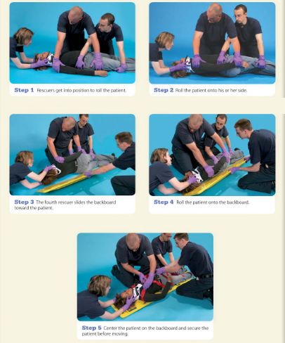
Log Rolling Maneuver : Steps
Step 1: Rescuer 1 stabilizes head and neck in neutral position without applying traction. He/she should grasp the patient’s shoulders at the neck and gently position the patient’s head betweeen the forearms. Another rescuer should apply a semirigid extrication collar. Even with the collar in place, Rescuer 1 must maintain…
Emergency Medicine
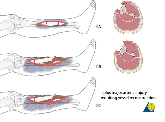
Open fractures : Mnemonics
Gustilo Anderson Classification Mnemonics:1. Parameters: ABCD’S (Area, Bone, Circulation, Dirt, Soft tissue)2. Classification: I, II, III then A, B, C Progression for grade I to III C implies a higher degree of energy involved in the injury, higher soft tissue and bone damage, and higher potential for complications. Type I:…