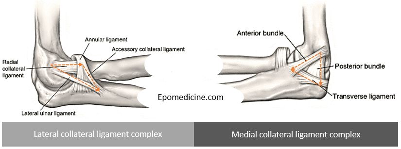
Lateral collateral ligament complex Restraint to varus and posterolateral rotatory instability. Anatomy is more variable. LCL arises from lateral humeral condyle at a point through which the axis of rotation passes – it maintains a uniform tension throughout the arc of motion. Annular ligament is a “U” shaped ligament that…
Tag: Musculoskeletal system
Emergency Medicine
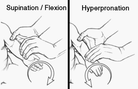
Pulled elbow reduction
Synonyms: Nursemaid’s elbow, Radial head subuluxation, Elbow subluxation Age: Commonly 1-4 years After 5 years of age, the attachment of the annular ligament to the neck of the radius strengthens Enlargement of the proximal radial epiphysis with growth may also improve stability Presentation: History of pull may be absent in…
PGMEE, MRCS, USMLE, MBBS, MD/MS
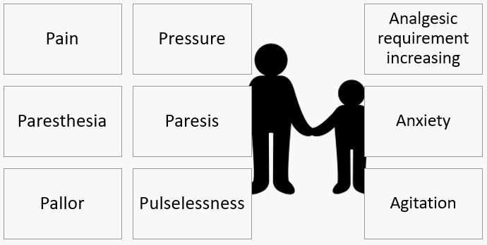
6 Ps and 3 As of Compartment Syndrome
Clinical features A. Adolescents and Adults Mnemonic: 6 Ps (by Hargens and Mubarak) Pain (may be absent in cases of nerve damage): Pain out of proportion to other physical findings (requiring increasing analgesic requirement) *: Earliest symptom Pain on passive stretch of the muscles in concerned compartment * Low sensitivity…
PGMEE, MRCS, USMLE, MBBS, MD/MS
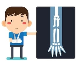
General Principles – Pediatric Fracture Management
Children are not just mini-adults • Higher head to torso ratio: Head injury & Upper C-spine fractures • Light weight – projectile when struck • BSA to Wt. ratio – higher (rapid hypothermia) • Large cardiopulmonary reserves – normal SBP in significant hypovolemia Growth contribution by Proximal & Distal Physes…
Emergency Medicine
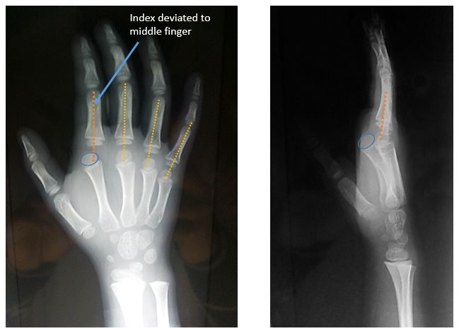
Kaplan’s Lesion : Complex Dorsal MCP joint dislocation
What is Kaplan’s lesion? Metacarpophalangeal (MCP) joint dislocation: Complex (Irreducible) Dorsal Mechanism of injury: Hyperextension injury Involvement: Usually occurs on border digits (Index > Little finger) Characteristic position of index finger: Hyperextended at MCP rests on dorsum of metacarpal Deviated towards middle finger Slightly flexed middle & distal phalanx with…
Clinical Skills and Approaches
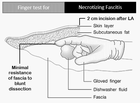
Finger test for Necrotizing Fascitis
Synonyms: Finger sweep test Finger test can be performed under Local anesthesia or General anesthesia in: Emergency department Bedside in wards Operation theaters Procedure: Area is infiltrated with local anesthetic A 2 cm test incision down to fascia is made in the suspected area The tissues are visually examined for:…
PGMEE, MRCS, USMLE, MBBS, MD/MS
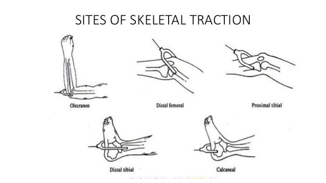
Skeletal Traction – Pin Insertion Sites
Site Point of insertion Direction Indications Olecranon (K-wire) 1.25 inches (3 cm) distal to olecranon tip – deep to subcutaneous border of upper ulna (avoids ulnar joint and open epiphysis) Medial to Lateral – At right angles to longitudinal axis of ulna (Avoids ulnar nerve) Supracondylar or Distal humerus fractures…
Clinical Skills and Approaches
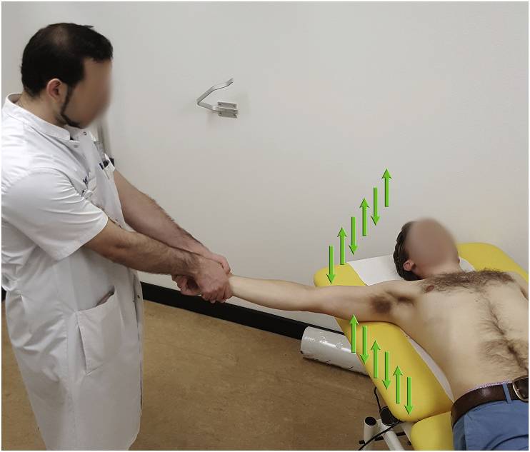
FARES method of Shoulder Reduction
Indication: Anterior shoulder dislocation Position of patient: Supine Position of physician: Standing on the side of the dislocated shoulder Steps: Physician holds the wrist of the patient with both the arms keeping elbow of the patient extended and forearm in neutral position Arm is slowly abducted in brief oscillating movement…