70% of blood supply of scaphoid is by dorsal branch of radial artery 70% of scaphoid bones have arterial foramina through out their length 70% of carpal fractures are scaphoid fractures 70% of scaphoid fractures occur through the waist 70% of scaphoid fractures are detected on initial radiographs 70% proximal…
Tag: Musculoskeletal system
PGMEE, MRCS, USMLE, MBBS, MD/MS
Pes Anserinus : Mnemonic
Pes Anserinus is composed of the combination of tendinous insertions of the sartorius, gracilis and semitendinosus muscles (guy ropes muscles) which attaches to the medial side of tibia to generate a “goose’s foot” like appearance. These three muscles are mainly flexors of the knee but also have a role in…
PGMEE, MRCS, USMLE, MBBS, MD/MS
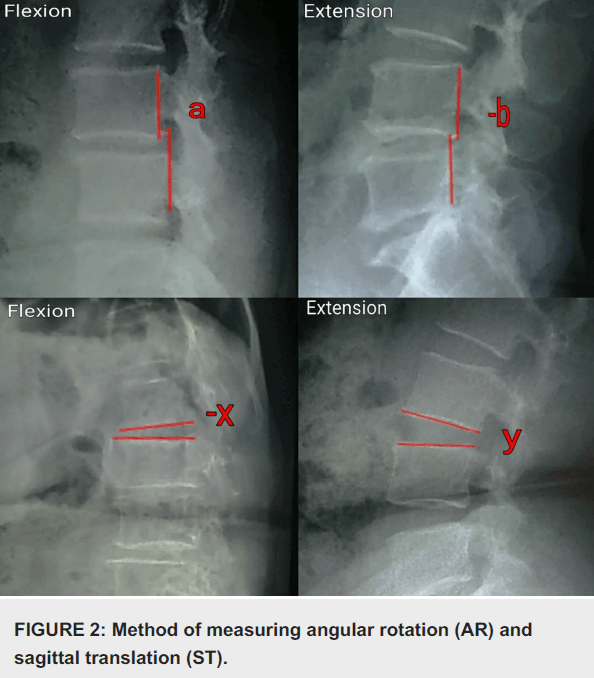
Diagnosis of Spinal Instability (White & Panjabi) : Mnemonic
Mnemonic: Remember 4 D common to all + 3 D for C-spine and 1 D for L-spine Diagnosis requires: Score >4 Denis column disruption: Anterior column destroyed or unable to function (2 points) Posterior column destroyed or unable to function (2 points) Deformity: Will have a maximum score of 4…
PGMEE, MRCS, USMLE, MBBS, MD/MS
Scheurmann’s Disease : Mnemonic
Mnemonic: Remember “S” for Scheurmann Structural kyphosis (sagittal deformity) of thoracic or thoracolumbar spine Skeletally immature sons (0.4-10% of adolescents between 10-14 years; onset with prepubertal growth spurt; M:F = 2-7:1) Strong hereditary (genetic) predisposition and Several theories: Scheurmann’s vertebral epiphyseal disturbance theory Schmorl’s nodes (herniation of disc material into…
PGMEE, MRCS, USMLE, MBBS, MD/MS
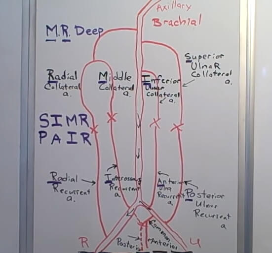
Elbow Anastomoses : Mnemonic
First things first. It is essential to understand the meaning of collateral and recurrent arteries. Recurrent arteries turn back so as to reverse direction. Collateral arteries refer to side branches of the major arteries. Mnemonic: M.R. Deep On the posterior aspect of the shaft of the humerus: Profunda brachii (Deep…
PGMEE, MRCS, USMLE, MBBS, MD/MS
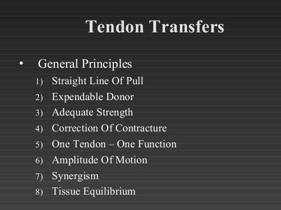
Tendon Transfer Principles : Mnemonic
Tendon transfer is the use of the power of a functioning muscle unit to activate a non-functioning nerve/muscle/tendon unit. The transferred tendon remains attached to its parent muscle with an intact neurovascular pedicle. Mnemonic: SEACOAST-1 a. Synergistic: act together to produce a single composite movement (facilitate each other). e.g. b….
PGMEE, MRCS, USMLE, MBBS, MD/MS
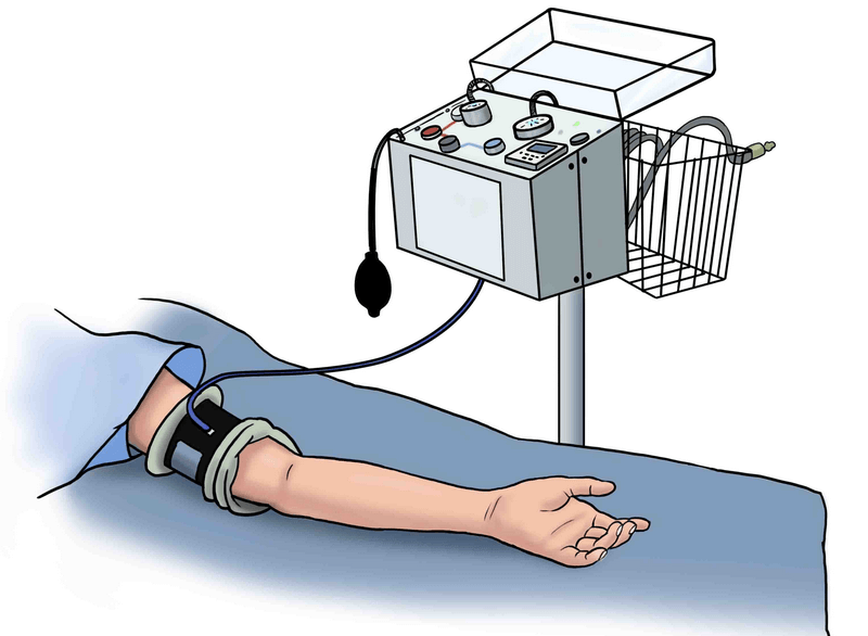
Tourniquet Paralysis Syndrome
Synonym: Pressure paralysis Mechanism: Direct extrinsic pressure (displacement of ranvier node) or axonal hypoxia on the nerves beneath the tourniquet and are related to the cuff pressure and duration of application.\ It is different from post tourniquet syndrome which caused due to combined effect of muscle ischemia, edema and microvascular…
PGMEE, MRCS, USMLE, MBBS, MD/MS
Blood Supply of Humeral Head
Anterior Humeral Circumflex Artery (AHCA) Origin: Axillary artery Course: Along the inferior border of subscapularis Gives anterolateral ascending branch which courses along lateral aspect of bicipital groove entering the humeral head and becoming arcuate artery Continues posterolaterally to anastomose with Posterior Humeral Circumflex Artery (PHCA) Posterior Humeral Circumflex Artery (PHCA)…