Acceptable angulation in Femoral Shaft Fractures
| Age | Varus/Valgus (degrees) | Anterior/Posterior (degrees) | Shortening (mm) |
| <2 yr | 30 | 30 | 15 |
| 2-5 yr | 15 | 20 | 20 |
| 6-10 yr | 10 | 15 | 15 |
| >10 yr | 5 | 10 | 10 |
Leg position for Hip Spica Application in Femur Shaft Fracture
Muscle forces:
- Proximal 1/3 fractures: Proximal fracture tends to be abducted and flexed by the insertion of muscles at the trochanters
- Midshaft fractures: Tend to fall in varus and external rotation (valgus mold is important)
- Distal 1/3 fractures: Gastrocnemius tends to pull the fracture into recruvatum (flexing the knee takes tension off the muscle and helps to maintain reduction)
| Location of fracture | Hip flexion | Hip abduction | Hip external rotation |
| Proximal 1/3 | 45 | 45 | 20 |
| Middle 1/3 | 30 | 30 | 10 |
| Distal 1/3 | 20 | 20 | 10 |
| Location of fracture | Hip flexion | Hip abduction | Hip external rotation |
| Proximal 1/3 | 45 | 30 | 20 |
| Middle 1/3 | 30 | 20 | 15 |
| Distal 1/3 | 20 | 20 | 15 |
For convenience and child positioning the knee is flexed to about 70°. This allows for comfort whilst lying and sitting.

Available via license: CC BY-NC 4.0
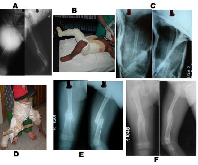
Ruhullah M, Singh HR, Shah S, Shrestha D. Primary hip spica with crossed retrograde intramedullary rush pins for the management of diaphyseal femur fractures in children: A prospective, randomized study. Niger Med J. 2014 Mar;55(2):111-5. doi: 10.4103/0300-1652.129638. PMID: 24791042; PMCID: PMC4003711.
More radical hip-knee flexion casts (so called 90-90) have commonly reported complications like nerve injury, skin slough or compartment syndrome. AO recommends against the use of 90-90 hip spica due to risk of compartment syndrome.1
Leg position for Hip spica application in Developmental Dysplasia of Hip
Human position (Salter position): Hip joint in 95° flexion and 40-45° abduction (best for maintaining hip stability and minimizing the risk of osteonecrosis)
“Safe zone” of Ramsey: Between maximum adduction before re-dislocation and excessive abduction, which increases risk of osteonecrosis.
Technique of Hip Spica Application
- Anesthesia: General anesthesia or conscious sedation
- Telescope test under fluoroscopy: shortening >3 cm is associated with excessive periosteal stripping and is associated with increased risk of loss of reduction in cast (consider flexible nails, external fixation or traction with delayed casting)
- Stockinette: Apply a stockinette or waterproof pantaloons cast liner
- Position: Position the patient on a spica-cast application table. Place a folded towel over the central abdomen, inside the tubular bandage, to create space in the cast for breathing.
- Position the leg as per indication (described above)
- Cotton padding: Overwrap the cast liner/stockinette with cotton padding from nipple line to ankle
- Extra-padding or felt:
- At the edges
- Knee
- Level of the right groin and carry it posteriorly across the gluteal fold, over the right iliac crest, in front of the abdomen, over the lateral aspect of the left thigh, and to the left inguinal area
- Cast: Apply fiberglass or plaster cast material starting 1 inch below the edge and ending 1 inch above the malleoli in 2 sections:
- Proximal section: Nipple line to knees
- Distal section: Knees to ankles
- Splints:
- Apply 4-5 plaster splints back to front from the nipple line to the back of the sacrum
- Apply a short, thick splint over the anterolateral aspect of the inguinal area
- Apply another splint starting from the right inguinal area, carry it posteriorly across the gluteal region, the iliac crest, the front of the abdomen, and back the same way on the opposite thigh
- Apply two splints over the medial and lateral aspects of the thigh, knee, and leg
- Mold for femur fractures: Apply an iliac crest mold to stabilize the hip and apply an anterior and valgus mold to the involved thigh to recreate the anterior bow and address the tendency of femoral shaft fractures to drift into varus.
- After care:
- Assess distal neurovascular status
- For fractures:
- Follow-up X-rays at 10 days after cast application:
- Unacceptable angulation – wedging
- Excessive shortening – reapplication of cast or change to another stabilization modality
- Cast removal – after 4 to 6 weeks
- Follow-up X-rays at 10 days after cast application:
- For DDH:
- Post-op: CT/USG/MRI
- After 6 wks:
- Remove 1st cast
- Stability assessed in moderate ROM
- AP pelvis radiograph
- Reduced 🡪 2nd cast
- Not reduced 🡪 Arthrogram
- After 12 wks: Remove 2nd cast 🡪 3rd cast X 6wks or Abduction splinting
References:
- Sargent MC. Single-Leg Spica Cast Application for Treatment of Pediatric Femoral Fracture. JBJS Essent Surg Tech. 2017 Sep 13;7(3):e26. doi: 10.2106/JBJS.ST.15.00070. PMID: 30233961; PMCID: PMC6132712.
- Hip spica casting for 32-D/5.1 Complete oblique or spiral, simple (aofoundation.org)
- Campbell’s Operative Orthopedics – 14th Edition
- Tachdjian’s Pediatric Orthopedics – 6th Edition

He is the section editor of Orthopedics in Epomedicine. He searches for and share simpler ways to make complicated medical topics simple. He also loves writing poetry, listening and playing music.