Position: Elbow flexed 90 degree and forearm in mid-prone
Landmarks:
- Lateral epicondyle of humerus
- Tip of radial styloid
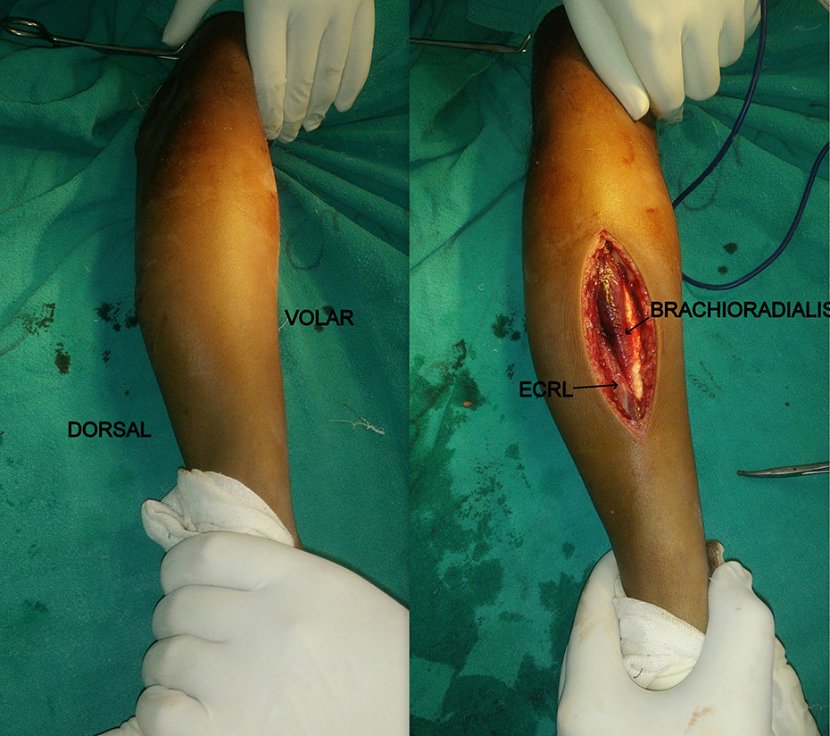
Skin incision: Centered over fracture in a straight imaginary line joining lateral epicondyle of humerus and tip of radial styloid
Superficial dissection: Subcutaneous tissue and deep fascia are incised along the line of skin incision
Deep dissection: Interval is developed between Brachioradialis and Extensor carpi radialis longus (ECRL)
- Superficial radial nerve is not isolated from brachioradialis unless it is crossing the surgical field
- Pronator teres can be divided partially or completely
Haseeb M, Muzafar K, Ghani A, Bhat KA, Butt MF. A fresh look at radial shaft fracture fixation: The lateral approach to the radius. Journal of Orthopaedic Surgery. 2018;26(2). doi:10.1177/2309499018780871 [CC BY-NC 4.0]
Fracture fixation:
- Plate is contoured according to the lateral bow of the radius and plate is applied on the lateral surface of the radius
Further reading:
- Haseeb M, Muzafar K, Ghani A, Bhat KA, Butt MF. A fresh look at radial shaft fracture fixation: The lateral approach to the radius. Journal of Orthopaedic Surgery. 2018;26(2). doi:10.1177/2309499018780871
- Devaraj B, Navaneethan A, Direct lateral approach to shaft of radius – a cadaver study. Indian J Orthop Surg 2017;3(2):181-183
- Muhammad Hanif, Muhammad Saeed Akhtar, Raza Elahi Rana, Shahzad: Direct Lateral Approach to Shaft of Radius. Journal of Pakistan Orthopaedic Association 2014;26 (3):11-14.

He is the section editor of Orthopedics in Epomedicine. He searches for and share simpler ways to make complicated medical topics simple. He also loves writing poetry, listening and playing music. He is currently pursuing Fellowship in Hip, Pelvi-acetabulum and Arthroplasty at B&B Hospital.