
While there are about 19 medical colleges currently running in Nepal and few more waiting for approval, there is no official body to rank them scientifically based on academic or other criterion. The Webometrics Ranking of World Universities is produced by Cybermetrics Lab (CCHS), a unit of the Spanish National Research…
PGMEE, MRCS, USMLE, MBBS, MD/MS
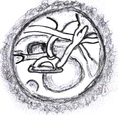
Anatomy of Middle Ear with Clinical correlation
The ear, along the evolution has modified structurally and functionally. In lower animals, they functioned as alarm systems to detect any sounds of the prey or predator so as to fix their vision and also maintain the balance of the body to prevent fall. In the course of evolution, this…
PGMEE, MRCS, USMLE, MBBS, MD/MS

Aplastic Anemia : Review notes
Definition Failure of bone marrow to produce peripheral blood cells and its progenitors Etiology The following illustration gives a brief idea about the etiological factors of aplastic anemia Now going into each etiological factor: – Autoimmune diseases: – Either they affect all the lineages (autoimmune aplastic anemia) or a single…
Emergency Medicine
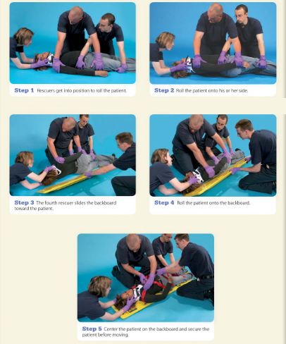
Log Rolling Maneuver : Steps
Step 1: Rescuer 1 stabilizes head and neck in neutral position without applying traction. He/she should grasp the patient’s shoulders at the neck and gently position the patient’s head betweeen the forearms. Another rescuer should apply a semirigid extrication collar. Even with the collar in place, Rescuer 1 must maintain…
PGMEE, MRCS, USMLE, MBBS, MD/MS
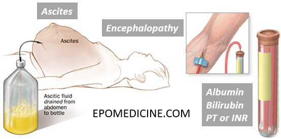
Hepatorenal syndrome (HRS) – Quick revision
New Criteria for HRS 1. Cirrhosis with ascites 2. Serum creatinine >1.5mg/dl 3. No sustained improvement in renal function after 2 days of diuretic withdrawl (if on diuretics) and volume expansion with albumin infusion at 1 gm/kg/day upto a maximum of 100 gm/day. 4. No evidence of shock 5. No…
Emergency Medicine
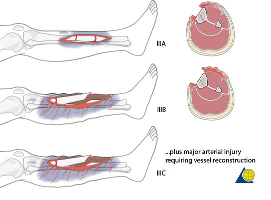
Open fractures : Mnemonics
Gustilo Anderson Classification Mnemonics:1. Parameters: ABCD’S (Area, Bone, Circulation, Dirt, Soft tissue)2. Classification: I, II, III then A, B, C Progression for grade I to III C implies a higher degree of energy involved in the injury, higher soft tissue and bone damage, and higher potential for complications. Type I:…
PGMEE, MRCS, USMLE, MBBS, MD/MS
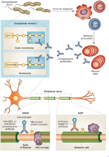
Guillain Barre Syndrome (GBS) – Mnemonic
ASBURY CRITERIA FOR GUILLAIN BARRE SYNDROME (GBS) Required Criteria Mnemonic: AIDP 1. Areflexia 2. Include in differential and rule out other causes 3. Duration < 4 weeks 4. Progressive weakness of 2 or more limbs due to neuropathy Supportive criteria Mnemonic: AIDPS 1. Afebrile 2. Involvement: Mild sensory involvement Facial…
Emergency Medicine
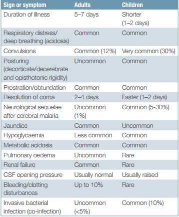
Severe Malaria : Quick revision
Criteria for Severe and Complicated Malaria Positive peripheral blood smear for P.falciparum + ≥1 of the CHAPLINS (Mnemonic) Convulsions: >2 in 24 hour Cerebral edema (Consciousness impaired) Hypoglycemia (glucose <40 mg/dl) Hemorrhage (DIC) Hemoglobinuria (Black water fever) Anemia (hemoglobin <5 gm/dl or PCV <15% in children; hemoglobin <7 gm/dl or…