Skip to content
Epomedicine

Mnemonics, Simplified Concepts & Thoughts

Epomedicine

Mnemonics, Simplified Concepts & Thoughts

Wormian Bones

Epomedicine, Aug 7, 2024Aug 7, 2024

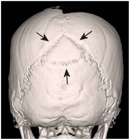
Wormian bones are abnormal ossicles that develop from extra ossification centers within the cranium. They are most frequently located in the lambdoid suture or the coronal suture, and have been seen in the fontanelles, particularly the posterior fontanelle.

wormian bone
Panzer S, Peschel O, Haas-Gebhard B, Bachmeier BE, Pusch CM, Nerlich AG, CC BY 2.5, via Wikimedia Commons

Commonest Cause

Idiopathic (Anatomic variant)

Other Causes

Mnemonic: PORK CHOPS

1. Pyknodyostosis

2. Osteogenesis imperfecta

3. Rickets

4. Kinky hair syndrome

5. Cleidocranial dysostosis

6. Hypothyroidism/hypophosphatasia

7. Otopalatodigital syndrome

8. Primary acro-osteolysis/Pachydermoperiostosis/Progeria

9. Syndrome of Downs

  • Facebook
  • Twitter
PGMEE, MRCS, USMLE, MBBS, MD/MS Musculoskeletal systemRadiology

Post navigation

Previous post
Next post

Related Posts

PGMEE, MRCS, USMLE, MBBS, MD/MS

Pathophysiology of Acute Compartment syndrome

Apr 27, 2023Apr 27, 2023

Definition Mubarak and Hargens (1981) defined compartment syndrome as an elevation of the interstitial pressure in a closed osseofascial compartment that results in microvascular compromise. Pathophysiology Tissue perfusion = Capillary perfusion pressure – Interstitial fluid pressure 3 major theories for microvascular dysfunction and ischemia: Tissue survival: Vicious cycle of compartment…

Read More
PGMEE, MRCS, USMLE, MBBS, MD/MS ddh lines angles

Lines and Angles in Orthopedics

Jun 9, 2019Jun 9, 2019

Slipped Capital Femoral Epiphysis (SCFE) Southwick’s angle: Epiphyseal-shaft angle on lateral radiograph <30 degrees: mild 30-60 degrees: moderate >60 degrees: severe Klein’s line: Line drawn along superior border of femoral neck (AP x-ray) In SCFE: will intersect less of the femoral head or not at all Developmental Dysplasia of Hip…

Read More
PGMEE, MRCS, USMLE, MBBS, MD/MS erythropoietin mechanism

Erythropoietin (EPO) Physiology

Sep 2, 2017

Structure of erythropoietin (EPO) Glycoprotein hormone 165 amino acids Molecular mass – 30 kDa Site of production/synthesis of erythropoietin (EPO) Kidneys (75-90%): Peritubular interstitial cells Liver (15%; chief source in fetus and neonates): Centrilobular hepatocytes After birth, erythropoietin is not detectable until 8-12 weeks after birth leading to physiological anaemia…

Read More

Leave a Reply Cancel reply

Your email address will not be published. Required fields are marked *

This site uses Akismet to reduce spam. Learn how your comment data is processed.

Pre-clinical (Basic Sciences)

Anatomy

Biochemistry

Community medicine (PSM)

Embryology

Microbiology

Pathology

Pharmacology

Physiology

Clinical Sciences

Anesthesia

Dermatology

Emergency medicine

Forensic

Internal medicine

Gynecology & Obstetrics

Oncology

Ophthalmology

Orthopedics

Otorhinolaryngology (ENT)

Pediatrics

Psychiatry

Radiology

Surgery

RSS Ask Epomedicine

  • What to study for Clinical examination in Orthopedics?
  • What is the mechanism of AVNRT?

Epomedicine weekly

  • About Epomedicine
  • Contact Us
  • Author Guidelines
  • Submit Article
  • Editorial Board
  • USMLE
  • MRCS
  • Thesis
©2026 Epomedicine | WordPress Theme by SuperbThemes