Before moving to the concept of wound closure, it is necessary to understand the pathophysiology behind different types of healing. We have covered stages of wound healing in general previously.
There are three types of wound healing:
- Primary intention
- Secondary intention
- Tertiary intention
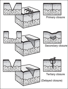
Healing by primary intention occurs when the dermal edges are approximated close together. Each suture track is a separate wound and incites the same phenomena as in healing by primary intention. There is minimal scarring is such type of wound healing.
Healing by secondary intention occurs when the dermal edges are not apposed with sutures but is left open. There is a larger tissue defect that needs to be bridged and hence, the healing occurs from the base upwards with proliferation of granulation tissue until it reaches the level of epithelium after which the epithelia can regenerate to re-epithelialize the gap. Contraction of wound to 1/3-1/4 of it’s original size by the action of myofibroblasts is an important feature of secondary intention healing which is not seen in that with primary intention. This type of healing is slow, has intense inflammatory phase and results in large, at times ugly scar.
Healing by tertiary intention occurs when the wound is left open initially and after a short period of time, wound edges are approximated and closed. It involves aspects of both primary and secondary wound healing. If the infection occurs at the site of surgical incision that is healing by primary intention, healing by tertiary intention may result due to an enlargement of injured area and increased in the magnitude and duration of inflammatory response.
Timing of Wound Closure
As with the intention of healing, there are three corresponding types of wound closure based on their timing of closure. Many surgeons mistaken tertiary closure with secondary closure.
Primary wound closure:
- Window of closure following injury: Within 4-12 hours for limbs and body and upto 24 hours for highly vascular areas like face and scalp (the proposed cut-off of golden period is highly variable in surgical textbooks and literatures)
- Indications: Simple, clean, non-contaminated wounds with minimal tissue loss and surgical incisions
- Mechanism: Healing by primary intention
Secondary wound closure:
- Wounds are not sutured to allow healing by secondary intention
- Indications: Skin infarctions, ulcerations, abscess cavities, punctures, small consmetically unimportant anatomical bites, and partial thickness (abrasions, second degree burns)
Delayed primary or Tertiary wound closure:
- Timing of closure: Beyond the time of golden period (4-24 hours as described earlier)
- Indications: Bite injuries, Infected injuries without significant tissue loss until infection control, Lacerations involving foreign body
- Mechanism: Healing by tertiary intention
Reference:
1. Wounds and Lacerations: Emergency Care and Closure By Alexander Trott
2. Healing by Intention, Advances in Skin & Wound Care: June 2017 – Volume 30 – Issue 6 – p 246-247 doi: 10.1097/01.ASW.0000516787.46060.b2
Your expaination is excellent