Skip to content
Epomedicine

Mnemonics, Simplified Concepts & Thoughts

Epomedicine

Mnemonics, Simplified Concepts & Thoughts

shoulder xray

Shoulder X-ray views

Epomedicine, Jan 2, 2021Aug 7, 2024

Shoulder X-ray views

AP Shoulder: in plane of thorax

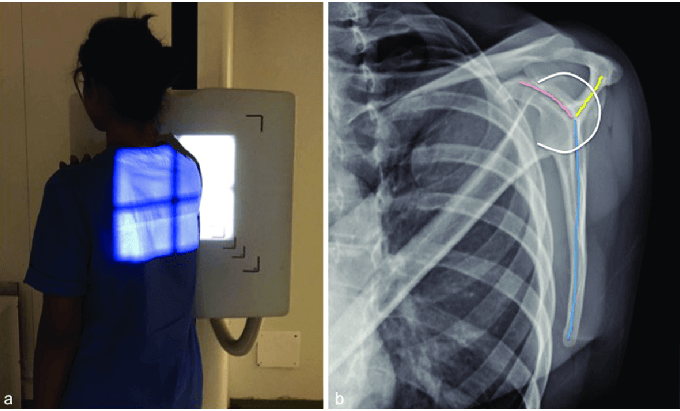
AP in plane of scapula: Angled 45 degrees lateral

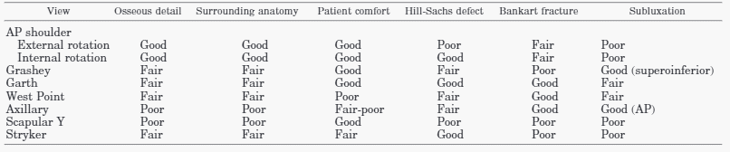
  • Neutral rotation: Grashey view (estimation of glenohumeral space)
  • Internal rotation/External rotation 30 degrees: Hill sach’s lesion and other humeral head morphology
grashey view
Grashey view

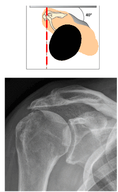
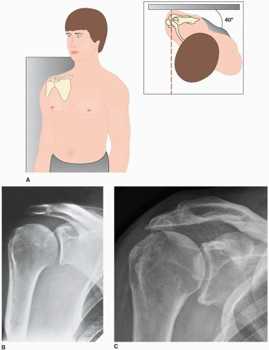
Scapular Y lateral: Erect with opposite shoulder rotated 40 degrees out and beam centered on spine of scapula (shoulder dislocations and scapula fractures)

scapular y view
Berritto, Daniela & Pinto, António & Russo, Anna & Urraro, Fabrizio & Laporta, Antonella & Belfiore, Maria Paola & Grassi, Roberto. (2018). Scapular fractures: A common diagnostic pitfall. Acta bio-medica : Atenei Parmensis. 89. 102-110. 10.23750/abm.v89i1-S.7014. [CC BY-NC-SA 4.0]

Supraspinatus outlet view: Scapular Y view with beam caudally tilted 10 degrees (Acromion morphology)

Axillary views:

  • Standard axillary (Lawrence): Supine; arm abducted 90 degrees; beam inferior to superior, 15 to 30 degrees medial angulation depending on abduction
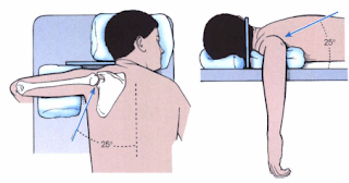
  • West Point axillary: Prone; arm abducted 90 degrees and hanging over the table edge; beam angulated 25 degrees medially and anteriorly (detect Bankart fractures)
  • Velpeau: Standing; wearing arm sling, leans backward over table 30 degrees; beam directed superior to inferior
  • Trauma axillary lateral: Supine; arm supported in 20 degrees flexion by placing radiolucent material under elbow
westpoint view
INFEROSUPERIOR AXIAL SHOULDER X-RAY – RadTechOnDuty [CC BY-NC-ND]

Apical Oblique (Garth view): Grashey (True AP) view with beam 45 degrees caudally (to show anterior/inferior capsule; bony bankart/Hill Sachs)

Garth view
INFEROSUPERIOR AXIAL SHOULDER X-RAY – RadTechOnDuty [CC BY-NC-ND]

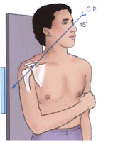
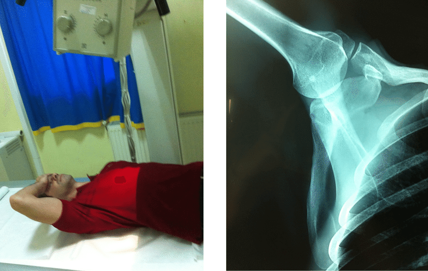
Stryker-notch view: Supine; palm on head with elbow straight up; beam directed AP with 10 degrees cephalic aiming coracoid (Hill Sachs)

Stryker notch view
Kostretzis, Lazaros & Theodoroudis, Ioannis & Boutsiadis, Achilleas & Papadakis, Nikolaos & Papadopoulos, Pericles. (2017). Suprascapular Nerve Pathology: A Review of the Literature. The Open Orthopaedics Journal. 11. 140-153. 10.2174/1874325001711010140. [CC BY 4.0]

Zanca view: Erect; Beam centered on AC joint with 10 degrees cephalic tilt and 50% exposure (AC Joint)

Serendipity view: Supine; Beam centered on clavicle/manubrium with 40 degrees cephalic tilt (SC Joint)

sternoclavicular joint views

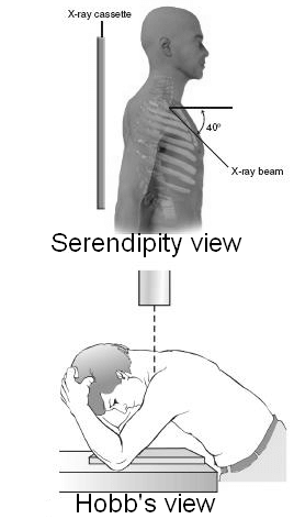
Hobb’s view (90 degree cephalocaudal lateral): Sitting; Leans forward so that the anterior chest is in contact with film cassette & the flexed elbows straddle the cassette & support the patient; X-ray beam is directed directly down


For specific pathologies

a. Obligatory: AP, Lateral or axillary

b. Subacromial impingement: “Y” view

c. Coracoid impingement: Axillary, Lateral

d. Scapular pathology: “Y” view

e. Bankart: Garth view, West point view

f. Hill Sach’s: Stryker notch view, AP with Internal rotation

g. Glenohumeral arthritis: True AP in internal and external rotation and Axillary lateral view

h. Sternoclavicular pathology: Serendipity view, Hobb’s view

i. Acromioclavicular pathology: Zanca view; Stress view with 5 kg weight suspended on wrist and showing both shoulders

  • Facebook
  • Twitter
PGMEE, MRCS, USMLE, MBBS, MD/MS Musculoskeletal systemOrthopedicsRadiology

Post navigation

Previous post
Next post

Related Posts

PGMEE, MRCS, USMLE, MBBS, MD/MS

Spinal segments vs Vertebral level

Aug 9, 2023Aug 9, 2023

Spinal segment: The part of spinal cord to which a pair of spinal nerves (right and left) is attached. Cause of mismatch between spinal segments and vertebral level: General rule: Vertebral level Add Spinal segment Cervical + 0 Same C7-T1 C8 T1-T3 +1 T1-T4 T3-T6 +2 T5-T8 T7-T8 +3 T9-T11…

Read More
PGMEE, MRCS, USMLE, MBBS, MD/MS

Spleen, Liver, Kidney Trauma

Jul 5, 2024Jul 5, 2024

Approach to abdominal trauma has been discussed earlier. Spleen Injury Liver Injury Kidney Injury General 2nd m/c injured intra-abdominal organ in blunt trauma m/c injury requiring operative intervention in blunt trauma m/c injured intra-abdominal organ overall m/c injured intra-abdominal organ in blunt trauma m/c from blunt trauma maximum warm ischemia…

Read More
PGMEE, MRCS, USMLE, MBBS, MD/MS

Wound Strength

Jul 5, 2025Jul 5, 2025

Wound strength is determined by both quantity and quality of collagen and an appropriate balance of deposition and breakdown. The amount of collagen in a wound plateaus at several weeks post-injury. Timeline: 1. First few weeks: Strength increases rapidly, reaching about 20% of pre-injury strength at 3 weeks.  2. Around 6 weeks: Wound strength…

Read More

Leave a Reply Cancel reply

Your email address will not be published. Required fields are marked *

This site uses Akismet to reduce spam. Learn how your comment data is processed.

Epomedicine. Shoulder X-ray views [Internet]. Epomedicine; 2021 Jan 2 [cited 2026 Jun 27]. Available from: https://epomedicine.com/medical-students/shoulder-x-ray-views/.

Pre-clinical (Basic Sciences)

Anatomy

Biochemistry

Community medicine (PSM)

Embryology

Microbiology

Pathology

Pharmacology

Physiology

Clinical Sciences

Anesthesia

Dermatology

Emergency medicine

Forensic

Internal medicine

Gynecology & Obstetrics

Oncology

Ophthalmology

Orthopedics

Otorhinolaryngology (ENT)

Pediatrics

Psychiatry

Radiology

Surgery

RSS Ask Epomedicine

  • What to study for Clinical examination in Orthopedics?
  • What is the mechanism of AVNRT?

Epomedicine weekly

  • About Epomedicine
  • Contact Us
  • Author Guidelines
  • Submit Article
  • Editorial Board
  • USMLE
  • MRCS
  • Thesis
©2026 Epomedicine | WordPress Theme by SuperbThemes