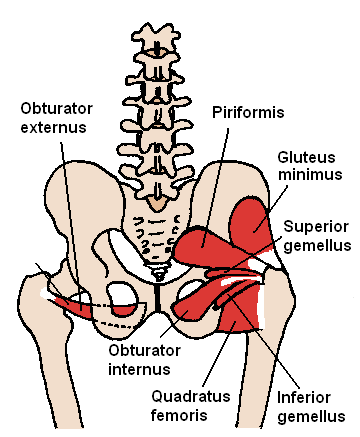
Short external rotators of hip in general originate from sacrum and ischium and insert on and around greater trochanter of femur.
Mnemonic: P GO GO Q
From proximal, the short external rotators of hip are:
- Piriformis
- Gemellus superior
- Obturator internus
- Gemellus inferior
- Obturator externus
- Quadratus femoris
| Muscle | Proximal attachment | Distal attachment | Innervation |
| Piriformis | Sacrum – anterior aspect Sacrotuberous ligament | Greater trochanter – superior aspect | Ventral rami S1, S2 |
| Gemellus superior | Ischial spine | Greater trochanter – medial aspect | Nerve to obturator internus (L5-S2) |
| Obturator internus | Internal surface of obturator membrane | Greater trochanter – medial aspect | Same as above |
| Gemellus inferior | Ischial tuberosity | Greater trochanter – medial aspect | Nerve to quadratus femoris (L4-S1) |
| Obturator externus | External surface of obturator membrane | Greater trochanter – medial aspect (trochanteric fossa) | Posterior branch of obturator nerve (L3, L4), i.e., lumbar plexus |
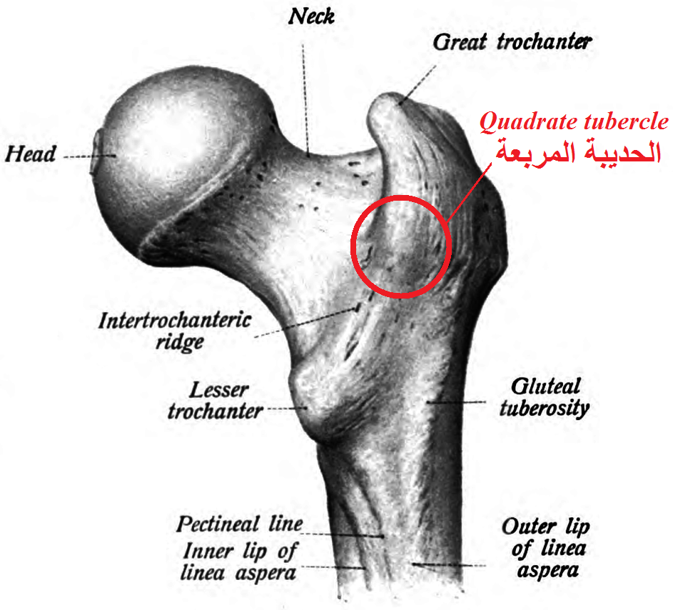
| Quadratus femoris | Ischial tuberosity – lateral border | Intertrochanteric crest – Quadrate tubercle | Nerve to quadratus femoris (L4-S1) |
Mnemonic: GSOI 512, IQ 451
Gemellus Superior – nerve to Obturator Internus (L5, S1, S2)
Gemellus Inferior – nerve to Quadratus femoris (L4, L5, S1)
Points of Surgical significance
1. Sciatic nerve emerges below piriformis and courses superficial to other short external rotators.
2. Branches of MFCA (Medial Femoral Circumflex Artery) emerge above and below Quadratus femoris.