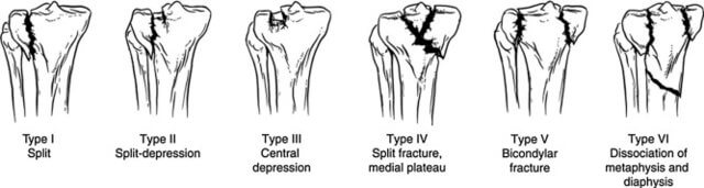
Schatzker system is widely accepted and used to classify tibial plateau fractures. It is based on a 2D representation of the fracture. As a general rule, it can be divided into low-energy and lateral tibial plateau variants (types I-III) and high energy variants (type IV-VI). However, it does not include fractures in the coronal plane or others not seen on plain AP radiographs. This classification helps separate the fractures into groups with similar mechanisms and patterns which will have similar treatment options.
Mnemonic: Imagine a break-up scenario in a failed romance and the various emotional stages you go through.
I – You have just split up and feel alone and on the outside.
II – Split and feeling depressed and still alone and on the outside.
III – Pure depression but still on the outside.
IV – You are on the otherside of the breakup and it doesn’t matter to you anymore about whether you are split or depressed.
V – You manage to see both sides of the split up.
VI – You have a relapse when you see him/her again and become a complete emotional mess.
Source: Thurston MD, Guyver PM, Jain NP, Toms AD. An aide-mémoire for tibial plateau fractures. Ann R Coll Surg Engl. 2014 Jul;96(5):386. doi: 10.1308/rcsann.2014.96.5.386. PMID: 24992425; PMCID: PMC4473938.
Now correlate this breakup story with the Schatzker classification.
Egypt J Radiol Nucl Med, 46 (3) (2015), pp. 695-699
CC BY-NC-ND 4.0
Schatzker originally defined depression as measuring greater than 4mm and as a surgical limit. The separation is defined as the diastasis between two fragments and the surgical limit is 2mm.
Generally, simple or incomplete fractures (Schatzker type 1) of the plateau are compressed with 6.5mm partially threaded cancellous screws. Complex type fractures will require a plate for enhanced stability.
I have devised a simpler mnemonic, which may be useful and help you remember the classiciation.
Mnemonic: SLeD MED
Type I – Split and Lateral
Type II – Split, Lateral and Depressed
Type III – Lateral and Depressed
Type IV – Medial
Type V – Either (Bicondylar)
Type VI – Diaphysis Dissociated from metaphysis
Additionally, the Hohl-Moore classification can be applied to fracture-dislocation variants that do not “fit” into the Schatzker classification.
- Type I – Coronal split
- Type II – Entire condylar
- Type III – Rim avulsion
- Type IV – Rim compression
- Type V – 4 part fracture

He is the section editor of Orthopedics in Epomedicine. He searches for and share simpler ways to make complicated medical topics simple. He also loves writing poetry, listening and playing music. He is currently pursuing Fellowship in Hip, Pelvi-acetabulum and Arthroplasty at B&B Hospital.
Schatzker II is a split-depression fracture and schatzker III is a pure depression. I think type II is more severe than type III. Is there any logic behind the classification?
The Schatzker classification, first described in 1979, is popular in North America. It combines fracture location and pattern and separately recognizes medial plateau fractures.
– SCHATZKER, JOSEPH*; MCBROOM, ROBERT**; BRUCE, DAVID† The Tibial Plateau Fracture: The Toronto Experience 1968–1975, Clinical Orthopaedics and Related Research: January-February 1979 – Volume – Issue 138 – p 94-104
May be the classification when formulated was not meant to denote the numerically increasing severity.
The only perspective which makes me think type III fractures as more severe than type II is that the direct visualization of the depressed articular surface is difficult in Schatzker type III fractures, even with a long anterior midline incision due to intact osseous rim. But I’m not sure about this.