Normal Rib Notching
A small notch near the costo-vertebral joint is normal, so pathologic rib notching is more likely if the notching is more lateral.
Types of Pathological Rib Notching
1. Superior rib notching
2. Inferior rib notching (more common)
Inferior Rib Notching (Roesler’s sign)
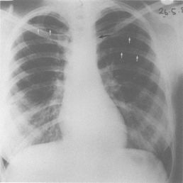
Mechanism: Enlargement of one or more of the structures that lie in the subcostal groove (intercostal nerve, artery and vein) – the reason that you were tought to not insert the needle inferior to the ribs during thoracocentesis.
Causes:
1. Arterial collaterals:
- Coarctation of aorta
- Takayasu arteritis or aortitis
- Blalock-Taussig shunt
- Arteriovenous malformation of chest
2. Venous collaterals:
- Superior venacava (SVC) obstruction
3. Neurogenic:
- Usually single: Schwannoma
- Usually multiple: Neurofibroma
Superior Rib Notching
Mechanism: Distrubance of osteoblastic and osteoclastic activity and the stress effect of intercostal muscle.
Causes:
1. Abnormal osteoblastic activity:
- Osteogenesis imperfecta
- Collagen vascular disorders: RA, SLE, Marfan syndrome, Sjogren’s syndrome
- Localized pressure effect: retractors in surgery, intercostal chest drainage
2. Abnormal osteoclastic activity: Hyperparathyroidism
3. Intercostal muscle stress:
- Paralytic poliomyelitis
- Restrictive lung disease
Both Inferior and Superior Notching
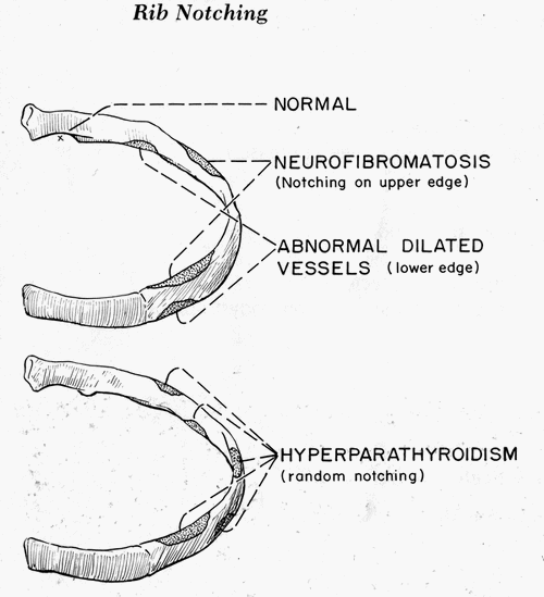
The diseases that can cause both superior and inferior rib notching are:
- Neurofibromatosis
- Hyperparathyroidism
- Coarctation of aorta with large collaterals
- Thalassemia
If both inferior and superior rib notching may be present, ribs may be called “ribbon ribs”.
Unilateral and Bilateral Rib Notching
Unilateral right sided inferior rib notching: coarctation at or proximal to the origin of left subclavian artery
Unilateral left sided inferior rib notching: coarctation proximal to an aberrant left subclavian artery
Other causes of unilateral inferior rib notching:
- Subclavian artery obstruction
- Blalock-Taussig shunt
Bilateral symmetrical inferior rib notching: coarctation distal to left subclavian artery
Bilateral asymmetrical and irregular inferior rib notching: neurofibromatosis
References: Fundamentals of Diagnostic Radiology edited by William E. Brant, Clyde A. Helms


Is blalocks operation associated with 4th and 5th rib notching
Most commonly after Blalock’s operation, unilateral rib notching of upper 3 or 4 ribs on upper side occurs.
Source: Aids to Radiological Differential Diagnosis By Stephen G. Davies, Stephen Chapman