Skip to content
Epomedicine

Mnemonics, Simplified Concepts & Thoughts

Epomedicine

Mnemonics, Simplified Concepts & Thoughts

Intrinsic thumb muscles : Tricks to Remember

Dr. Sulabh Kumar Shrestha, MS Orthopedics, Apr 26, 2020Oct 27, 2022

The muscles of thumb will make 2 compartments – thenar compartment and adductor compartment.

intrinsic thumb muscles

Mnemonic: Do it yourself as shown in the image below. Put your left hand with curled in middle/long finger above the palm of right hand. There are 2 muscle layers in this side of the hand. Represent the superficial layer with T-O-F-A (Transverse head, Oblique head, Flexor pollicis brevis, Abductor pollicis brevis) and the deeper later with T-O-F-O (Transverse head, Oblique head, Flexor pollicis brevis, Opponens pollicis). Curled in long finger represents the tendon of Flexor Pollicis LONGUS in the deeper layer between the OBLIQUE head of adductor pollicis and FLEXOR pollicis brevis.

The left little and ring finger on the ulnar side of right thumb represents 2 heads of adductor pollicis which forms the Adductor compartment.

Little finger (T-T): Transverse head of Adductor pollicis (in both superficial and deep layer)

  • As in the image below – it will originate from 3rd metacarpal and insert into ulnar side of base of proximal phalanx of thumb.

Ring finger (O-O): Oblique head of Adductor pollicis (in both superficial and deep layer)

  • This will originate from 3rd metacarpal as well as 2nd metacarpal and capitate and insert into ulnar base of proximal thumb phalanx like the transverse head.
thenar muscle mnemonic

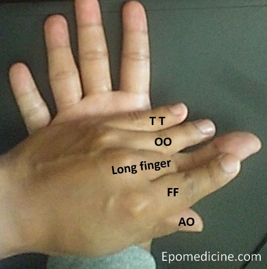
Now moving to the radial side of the thumb. Here, the muscles are different in different layers. All these muscles make Thenar compartment.

Index finger (F-F):

  • Superficial layer: FLEXOR pollicis brevis – superifical head (arises from flexor retinaculum and trapedzoid tubercle)
  • Deep layer: FLEXOR pollicis brevis – deep head (arises from trapezoid)

Both the heads, insert into radial base of proximal phalanx of thumb.

Thumb (A-O):

  • Superficial layer: ABDUCTOR pollicis brevis – originates from scaphoid and trapezium and insert into radial base of proximal phalanx of thumb.
  • Deep layer: OPPONENS pollicis – originates from trapezium and insert into lateral surface of 1st metacarpal shaft.

Innervation: The thenar muscles are supplied by motor recurrent branch of median nerve. The adductor muscle is supplied by ulnar nerve. The flexor pollicis brevis has dual innervation. While the superficial head is supplied by the median nerve, deep head receives it’s innervation from ulnar nerve.

dr. sulabh kumar shrestha
Dr. Sulabh Kumar Shrestha, MS Orthopedics

He is the section editor of Orthopedics in Epomedicine. He searches for and share simpler ways to make complicated medical topics simple. He also loves writing poetry, listening and playing music. He is currently pursuing Fellowship in Hip, Pelvi-acetabulum and Arthroplasty at B&B Hospital.

51 shares
  • Facebook51
  • Twitter
PGMEE, MRCS, USMLE, MBBS, MD/MS AnatomyMusculoskeletal system

Post navigation

Previous post
Next post

Related Posts

PGMEE, MRCS, USMLE, MBBS, MD/MS

Obstacles to reduction in Developmental Dysplasia of Hip (DDH)

Nov 25, 2025Mar 23, 2026

Extra-articular obstacles: Secondary muscle shortening due to hip in subluxed/dislocated position Intra-articular obstacles: Results in decreased volume of the acetabulum Dr. Sulabh Kumar Shrestha, MS Orthopedics He is the section editor of Orthopedics in Epomedicine. He searches for and share simpler ways to make complicated medical topics simple. He also…

Read More

Applied Anatomy of Tympanic Membrane

Nov 16, 2013May 11, 2024

Synonyms: Ear drum, Myringa Definition: Tympanic membrane is a thin membrane that separates the external ear from the middle ear. Anatomy: Site: Located at medial end of the external auditory canal, separating it from the middle ear cavity Position: At an angle of 55° to the horizontal to the floor…

Read More
PGMEE, MRCS, USMLE, MBBS, MD/MS

Mnemonic Approach to Elbow Xray – FOOL

Oct 12, 2019Jun 14, 2022

Mnemonic: FOOLa. Fat padsb. Overt findings and outlinesc. Ossification centersd. Lines Fat pads The elbow joint has anterior and posterior fat pads which are extra-synovial but within the articular capsule. These are present as a radiolucent (black) line (compared to bone and surrounding soft tissues) in lateral X-rays. Normal finding:…

Read More

Leave a Reply Cancel reply

Your email address will not be published. Required fields are marked *

This site uses Akismet to reduce spam. Learn how your comment data is processed.

Pre-clinical (Basic Sciences)

Anatomy

Biochemistry

Community medicine (PSM)

Embryology

Microbiology

Pathology

Pharmacology

Physiology

Clinical Sciences

Anesthesia

Dermatology

Emergency medicine

Forensic

Internal medicine

Gynecology & Obstetrics

Oncology

Ophthalmology

Orthopedics

Otorhinolaryngology (ENT)

Pediatrics

Psychiatry

Radiology

Surgery

RSS Ask Epomedicine

  • What to study for Clinical examination in Orthopedics?
  • What is the mechanism of AVNRT?

Epomedicine weekly

  • About Epomedicine
  • Contact Us
  • Author Guidelines
  • Submit Article
  • Editorial Board
  • USMLE
  • MRCS
  • Thesis
©2026 Epomedicine | WordPress Theme by SuperbThemes