Skip to content
Epomedicine

Mnemonics, Simplified Concepts & Thoughts

Epomedicine

Mnemonics, Simplified Concepts & Thoughts

Foot muscles – Layers and Compartments

Epomedicine, Sep 3, 2020Sep 3, 2020

Deep to the plantar fascia, muscles of plantar foot exist in 4 different layers. Extensor digitorum brevis makes dorsal layer of foot and remaining 18 muscles and 4 tendons make the 4 layers of plantar aspect or sole of foot.

In general:

  • Layer 1 and 3: 3 muscles each that contains intrinisc abductors, flexors and adductors
  • Layer 2 and 4: 2 muscles and 2 tendons that contains lumbricals and interossei

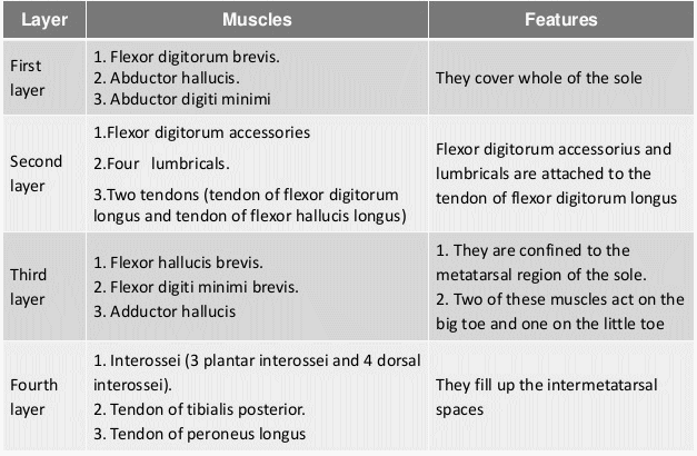
From superficial to deep, the layers are:

a. First layer

Mnemonic: Abs Flex Abs

  • Abductor hallucis
  • Flexor digitorum brevis (comparable to FDS of hand – both are superficial and insert into middle phalanx)
  • Abductor digiti minimii

b. Second layer

Mnemonic: Quadratus Lumborum OR you can also remember it with Flexor digitorum longus (comparable to FDP of hand – deeper and attaches to distal phalanx and the 2 muscles that attaches to FDL)

  • Quadratus plantae (Flexor digitorum accessorius)
  • Lumbricals (4)

Both of these muscles have attachment to the Flexor Digitorum Longus (FDL).

It also has 2 tendons of extrinsic muscles: Flexor digitorum longus (FDL) and Flexor hallucis longus (FHL).

It also has medial and lateral plantar nervers which are the terminal branches of tibial nerve.

c. Third layer

Mnemonic: Flex Add Flex (Opposite of first layer)

  • Flexor hallucis brevis
  • Adductor hallucis (like in hand, it has 2 heads – transverse and oblique)
  • Flexor digiti minimi

Both the flexor hallucis longus and brevis muscles have lateral origin, i.e. (distal fibula and cuboid/lateral cuneiform respectively) although they insert medially (towards great toe).

d. Fourth layer

Mnemonic: 4DAB and 3 PAD

  • 4 Dorsal interossei: ABduct MTP joint
  • 3 Plantar interossei: ADduct MTP joint
  • 2 tendons of extrinsic muscles: Peroneus longus and Tibialis posterior

9 compartments of foot

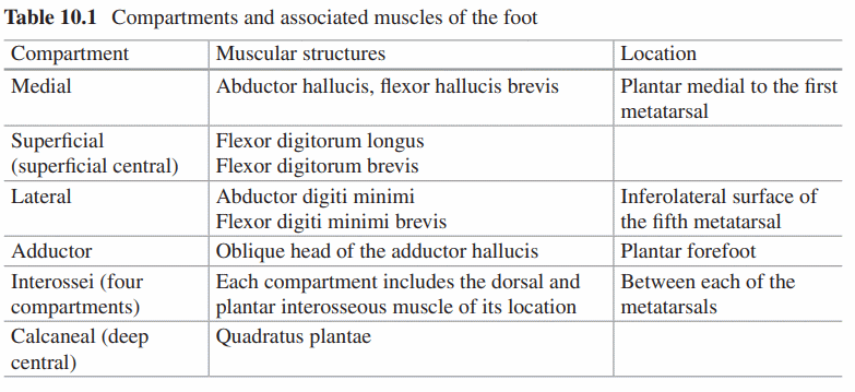
Please refer to an anatomy atlas and visualize for better understanding.

16 shares
  • Facebook
  • Twitter
PGMEE, MRCS, USMLE, MBBS, MD/MS AnatomyMusculoskeletal systemOrthopedics

Post navigation

Previous post
Next post

Related Posts

PGMEE, MRCS, USMLE, MBBS, MD/MS gastric bed

Surgical Anatomy of Stomach

Jan 5, 2016Feb 28, 2018

GASTROESOPHAGEAL JUNCTION (CARDIA) It is the junction between esophagus and cardia of stomach Histologically: Mucosal transition from squamous to columnar epithelium Functionally: High pressure zone (Lower esophageal sphincter or LES) – Normally, LES is intraperitoneal, >2 cm long, and has a resting pressure >6 mmHg; not an anatomical sphincter but a…

Read More
PGMEE, MRCS, USMLE, MBBS, MD/MS primary bone tumors and tumor-like lesions

Differential Diagnoses of Bone Tumors : Mnemonic Approach

May 31, 2019Oct 6, 2021

Lytic and Sclerotic Bone Lesions Mnemonic for Solitary lytic lesions: FEGNOMASHIC Fibrous dysplasia Enchondroma Eosinophilic granuloma Giant cell tumor Nonossifying fibroma Osteoblastoma, Osteosarcoma (telangiectatic) Metastases Myeloma Aneursymal bone cyst Simple bone cyst Hyperparathyroidism (brown tumor) Infection Chondroblastoma Chondromyxoid fibroma Mnemonic for Multiple lytic lesions: FEMHI Fibrous dysplasia Eosinophilic granuloma/Enchondroma Metastasis/Mutiple…

Read More
PGMEE, MRCS, USMLE, MBBS, MD/MS tertiary villi

Embryology Week 3: Trimalinar Germ Disc

Aug 3, 2016Aug 3, 2016

In the Week 2, we had a bilaminar germ disc with 2 cavities (amniotic cavity and yolk sac) suspended in the chorionic (extraembryonic cavity) by a connecting stalk (future umbilical cord). In the 3rd week, the embryo will have 3 germ layers (ectoderm, mesoderm and endoderm) following a process called…

Read More

Leave a Reply Cancel reply

Your email address will not be published. Required fields are marked *

This site uses Akismet to reduce spam. Learn how your comment data is processed.

Pre-clinical (Basic Sciences)

Anatomy

Biochemistry

Community medicine (PSM)

Embryology

Microbiology

Pathology

Pharmacology

Physiology

Clinical Sciences

Anesthesia

Dermatology

Emergency medicine

Forensic

Internal medicine

Gynecology & Obstetrics

Oncology

Ophthalmology

Orthopedics

Otorhinolaryngology (ENT)

Pediatrics

Psychiatry

Radiology

Surgery

RSS Ask Epomedicine

  • What to study for Clinical examination in Orthopedics?
  • What is the mechanism of AVNRT?

Epomedicine weekly

Epomedicine. Foot muscles – Layers and Compartments [Internet]. Epomedicine; 2020 Sep 3 [cited 2025 Nov 19]. Available from: https://epomedicine.com/medical-students/foot-muscles-layers-compartments/.

  • About Epomedicine
  • Contact Us
  • Author Guidelines
  • Submit Article
  • Editorial Board
  • USMLE
  • MRCS
  • Thesis
©2025 Epomedicine . All rights reserved.